 Movie
Movie Controller

[English] 日本語
 Yorodumi
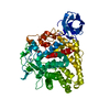
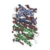
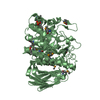
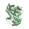
- PDB-1h80: 1,3-ALPHA-1,4-BETA-D-GALACTOSE-4-SULFATE- 3,6-ANHYDRO-D-GALACTOSE... -
+ Open data

- Basic information
| Entry | Database: PDB / ID: 1h80 | ||||||
|---|---|---|---|---|---|---|---|
| Title | 1,3-ALPHA-1,4-BETA-D-GALACTOSE-4-SULFATE- 3,6-ANHYDRO-D-GALACTOSE-2-SULFATE 4 GALACTOHYDROLASE | ||||||
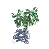
 Components Components | IOTA-CARRAGEENASE | ||||||
 Keywords Keywords | HYDROLASE / IOTA-CARRAGEENAN DOUBLE HELIX DEGRADATION | ||||||
| Function / homology |  Function and homology information Function and homology informationiota-carrageenase / iota-carrageenase activity / polysaccharide catabolic process / cell wall organization / extracellular region Similarity search - Function | ||||||
| Biological species |  ALTEROMONAS SP. ATCC43554 (bacteria) ALTEROMONAS SP. ATCC43554 (bacteria) | ||||||
| Method |  X-RAY DIFFRACTION / SYNCHROTRON / MAD / Resolution: 1.6 Å X-RAY DIFFRACTION / SYNCHROTRON / MAD / Resolution: 1.6 Å | ||||||
 Authors Authors | Michel, G. / Chantalat, L. / Dideberg, O. | ||||||
 Citation Citation | Journal: J.Biol.Chem. / Year: 2001 Title: The Iota-Carrageenase of Alteromonas Fortis. A Beta-Helix Fold-Containing Enzyme for the Degradation of a Highly Polyanionic Polysaccharide Authors: Michel, G. / Chantalat, L. / Fanchon, E. / Henrissat, B. / Kloareg, B. / Dideberg, O. #1: Journal: Acta Crystallogr.,Sect.D / Year: 2000 Title: Expression, Purification, Cristallization and Preliminary X-Ray Analysis of the Iota-Carrageenase from Alteromonas Fortis Authors: Michel, G. / Flament, D. / Barbeyron, T. / Vernet, T. / Kloareg, B. / Dideberg, O. | ||||||
| History |
|
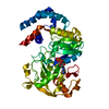
- Structure visualization
| Structure viewer | Molecule: MolmilJmol/JSmol |
|---|
- Downloads & links
-Download
| PDBx/mmCIF format | 1h80.cif.gz | 214.1 KB | Display | PDBx/mmCIF format |
|---|---|---|---|---|
| PDB format | pdb1h80.ent.gz | 167.8 KB | Display | PDB format |
| PDBx/mmJSON format | 1h80.json.gz | Tree view | PDBx/mmJSON format | |
| Others |  Other downloads Other downloads |
-Validation report
| Arichive directory | https://data.pdbj.org/pub/pdb/validation_reports/h8/1h80ftp://data.pdbj.org/pub/pdb/validation_reports/h8/1h80 | HTTPS FTP |
|---|
-Related structure data
| Related structure data | |
|---|---|
| Similar structure data |


-Links
 PDBj
PDBj- Assembly
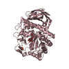
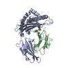
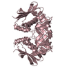
| Deposited unit | 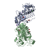
| ||||||||
|---|---|---|---|---|---|---|---|---|---|
| 1 | 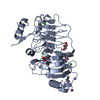
| ||||||||
| 2 | 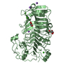
| ||||||||
| Unit cell |
| ||||||||
| Noncrystallographic symmetry (NCS) | NCS oper: (Code: given Matrix: (0.9928, 0.08692, 0.08239), Vector: |
-Components
-Protein , 1 types, 2 molecules AB
| #1: Protein | Mass: 51936.082 Da / Num. of mol.: 2 / Fragment: CATALYTIC DOMAIN RESIDUES 28-491 Source method: isolated from a genetically manipulated source Details: COMPLEXED WITH CALCIUM, SODIUM AND CHLORIDE / Source: (gene. exp.) ALTEROMONAS SP. ATCC43554 (bacteria) / Description: HIS-TAG, SELEMETHIONYL PROTEIN. / Gene: CGIA / Plasmid: PET20B / Production host: |
|---|
-Non-polymers , 5 types, 1130 molecules 


| #2: Chemical | ChemComp-CA /  Mass: 40.078 Da / Num. of mol.: 6 / Source method: obtained synthetically / Formula: Ca Mass: 40.078 Da / Num. of mol.: 6 / Source method: obtained synthetically / Formula: Ca#3: Chemical |  Mass: 35.453 Da / Num. of mol.: 2 / Source method: obtained synthetically / Formula: Cl Mass: 35.453 Da / Num. of mol.: 2 / Source method: obtained synthetically / Formula: Cl#4: Chemical | ChemComp-NA /  Mass: 22.990 Da / Num. of mol.: 6 / Source method: obtained synthetically / Formula: Na Mass: 22.990 Da / Num. of mol.: 6 / Source method: obtained synthetically / Formula: Na#5: Chemical | ChemComp-GOL /  Mass: 92.094 Da / Num. of mol.: 6 / Source method: obtained synthetically / Formula: C3H8O3 Mass: 92.094 Da / Num. of mol.: 6 / Source method: obtained synthetically / Formula: C3H8O3#6: Water | ChemComp-HOH / | Mass: 18.015 Da / Num. of mol.: 1110 / Source method: isolated from a natural source / Formula: H2O |
|---|
-Details
| Has protein modification | Y |
|---|
-Experimental details
-Experiment
| Experiment | Method: X-RAY DIFFRACTION / Number of used crystals: 1 |
|---|
- Sample preparation
| Crystal | Density Matthews: 3 Å3/Da / Density % sol: 52 % / Description: MAD DATA COLLECTION ON BM30 | |||||||||||||||||||||||||||||||||||
|---|---|---|---|---|---|---|---|---|---|---|---|---|---|---|---|---|---|---|---|---|---|---|---|---|---|---|---|---|---|---|---|---|---|---|---|---|
| Crystal grow | pH: 6.5 Details: 0.1 M SODIUM CACODYLATE PH 6.5, 10% GLYCEROL, 15-17% PEG6000, 200MM CALCIUM ACETATE | |||||||||||||||||||||||||||||||||||
| Crystal grow | *PLUS Temperature: 288 K / Method: vapor diffusion, hanging dropDetails: Michel, G., (2000) Acta Crystallogr.,Sect.D, 56, 766. | |||||||||||||||||||||||||||||||||||
| Components of the solutions | *PLUS
|
-Data collection
| Diffraction | Mean temperature: 100 K |
|---|---|
| Diffraction source | Source: SYNCHROTRON / Site: ESRF  / Beamline: ID14-2 / Wavelength: 0.933 / Beamline: ID14-2 / Wavelength: 0.933 |
| Detector | Type: MARRESEARCH / Detector: CCD / Date: Jul 15, 1998 / Details: MIRRORS |
| Radiation | Protocol: SINGLE WAVELENGTH / Monochromatic (M) / Laue (L): M / Scattering type: x-ray |
| Radiation wavelength | Wavelength: 0.933 Å / Relative weight: 1 |
| Reflection | Resolution: 1.6→20 Å / Num. obs: 762046 / % possible obs: 97.7 % / Redundancy: 4.8 % / Biso Wilson estimate: 18.4 Å2 / Rsym value: 0.123 |
| Reflection shell | Resolution: 1.6→1.66 Å / Rsym value: 0.203 / % possible all: 96.1 |
| Reflection | *PLUS Highest resolution: 1.6 Å / Redundancy: 4.8 % / Num. measured all: 157467 / Rmerge(I) obs: 0.123 |
| Reflection shell | *PLUS Highest resolution: 1.6 Å / % possible obs: 96.1 % / Rmerge(I) obs: 0.203 |
- Processing
| Software |
| ||||||||||||||||||||||||||||||||||||||||||||||||||||||||||||||||||||||||||||||||
|---|---|---|---|---|---|---|---|---|---|---|---|---|---|---|---|---|---|---|---|---|---|---|---|---|---|---|---|---|---|---|---|---|---|---|---|---|---|---|---|---|---|---|---|---|---|---|---|---|---|---|---|---|---|---|---|---|---|---|---|---|---|---|---|---|---|---|---|---|---|---|---|---|---|---|---|---|---|---|---|---|---|
| Refinement | Method to determine structure: MAD / Resolution: 1.6→20 Å / Rfactor Rfree error: 0.003 / Data cutoff high absF: 1960016.64 / Isotropic thermal model: RESTRAINED / Cross valid method: THROUGHOUT / σ(F): 0 Details: DOMAIN A HAS A HIGHER B FACTOR, REGIONS 314 - 334 AND 341 - 350 ARE NOT VISIBLE IN THE ELECTRON DENSITY MAP
| ||||||||||||||||||||||||||||||||||||||||||||||||||||||||||||||||||||||||||||||||
| Solvent computation | Solvent model: FLAT MODEL / Bsol: 50.2073 Å2 / ksol: 0.415064 e/Å3 | ||||||||||||||||||||||||||||||||||||||||||||||||||||||||||||||||||||||||||||||||
| Displacement parameters | Biso mean: 22.4 Å2
| ||||||||||||||||||||||||||||||||||||||||||||||||||||||||||||||||||||||||||||||||
| Refine analyze |
| ||||||||||||||||||||||||||||||||||||||||||||||||||||||||||||||||||||||||||||||||
| Refinement step | Cycle: LAST / Resolution: 1.6→20 Å
| ||||||||||||||||||||||||||||||||||||||||||||||||||||||||||||||||||||||||||||||||
| Refine LS restraints |
| ||||||||||||||||||||||||||||||||||||||||||||||||||||||||||||||||||||||||||||||||
| LS refinement shell | Resolution: 1.6→1.7 Å / Rfactor Rfree error: 0.007 / Total num. of bins used: 6
| ||||||||||||||||||||||||||||||||||||||||||||||||||||||||||||||||||||||||||||||||
| Xplor file |
| ||||||||||||||||||||||||||||||||||||||||||||||||||||||||||||||||||||||||||||||||
| Refinement | *PLUS Highest resolution: 1.6 Å / Lowest resolution: 20 Å / % reflection Rfree: 5 % | ||||||||||||||||||||||||||||||||||||||||||||||||||||||||||||||||||||||||||||||||
| Solvent computation | *PLUS | ||||||||||||||||||||||||||||||||||||||||||||||||||||||||||||||||||||||||||||||||
| Displacement parameters | *PLUS | ||||||||||||||||||||||||||||||||||||||||||||||||||||||||||||||||||||||||||||||||
| Refine LS restraints | *PLUS
| ||||||||||||||||||||||||||||||||||||||||||||||||||||||||||||||||||||||||||||||||
| LS refinement shell | *PLUS Lowest resolution: 1.66 Å |
