 Movie
Movie Controller

[English] 日本語
 Yorodumi
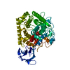
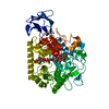
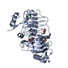
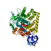
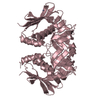
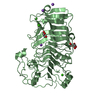
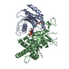
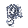
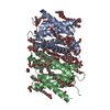
- PDB-3rx7: Structure of AaCel9A in complex with cellotetraose-like isofagomine -
+ Open data

- Basic information
| Entry | Database: PDB / ID: 3rx7 | |||||||||
|---|---|---|---|---|---|---|---|---|---|---|
| Title | Structure of AaCel9A in complex with cellotetraose-like isofagomine | |||||||||
 Components Components | Cellulase | |||||||||
 Keywords Keywords | HYDROLASE/HYDROLASE INHIBITOR / GH9 family / endoglucanase / HYDROLASE-HYDROLASE INHIBITOR complex | |||||||||
| Function / homology |  Function and homology information Function and homology informationcellulase / cellulase activity / cellulose catabolic process / metal ion binding Similarity search - Function | |||||||||
| Biological species |  Alicyclobacillus acidocaldarius subsp. acidocaldarius (bacteria) Alicyclobacillus acidocaldarius subsp. acidocaldarius (bacteria) | |||||||||
| Method |  X-RAY DIFFRACTION / SYNCHROTRON / MOLECULAR REPLACEMENT / Resolution: 2.02 Å X-RAY DIFFRACTION / SYNCHROTRON / MOLECULAR REPLACEMENT / Resolution: 2.02 Å | |||||||||
 Authors Authors | Morera, S. | |||||||||
 Citation Citation | Journal: Org.Biomol.Chem. / Year: 2011 Title: Fortuitious binding of inhibitors-derived isofagomine for inverting GH9 beta-glycosidases Authors: Morera, S. / Vigouroux, A. / Stubbs, K.A. | |||||||||
| History |
|
- Structure visualization
| Structure viewer | Molecule: MolmilJmol/JSmol |
|---|
- Downloads & links
-Download
| PDBx/mmCIF format | 3rx7.cif.gz | 121.3 KB | Display | PDBx/mmCIF format |
|---|---|---|---|---|
| PDB format | pdb3rx7.ent.gz | 91.9 KB | Display | PDB format |
| PDBx/mmJSON format | 3rx7.json.gz | Tree view | PDBx/mmJSON format | |
| Others |  Other downloads Other downloads |
-Validation report
| Arichive directory | https://data.pdbj.org/pub/pdb/validation_reports/rx/3rx7ftp://data.pdbj.org/pub/pdb/validation_reports/rx/3rx7 | HTTPS FTP |
|---|
-Related structure data
| Related structure data |  3rx5C  3rx8C  3gzkS S: Starting model for refinement C: citing same article ( |
|---|---|
| Similar structure data |


-Links
 PDBj
PDBj
- Assembly
| Deposited unit | 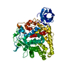
| ||||||||
|---|---|---|---|---|---|---|---|---|---|
| 1 |
| ||||||||
| Unit cell |
|
-Components
-Protein , 1 types, 1 molecules A
| #1: Protein | Mass: 59030.992 Da / Num. of mol.: 1 Source method: isolated from a genetically manipulated source Source: (gene. exp.) Alicyclobacillus acidocaldarius subsp. acidocaldarius (bacteria)Gene: celA / Production host: |
|---|
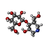
-Sugars , 2 types, 2 molecules 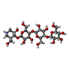
| #4: Sugar | ChemComp-G3I / ( Type: saccharide / Mass: 633.594 Da / Num. of mol.: 1 Type: saccharide / Mass: 633.594 Da / Num. of mol.: 1Source method: isolated from a genetically manipulated source Formula: C24H43NO18 |
|---|---|
| #5: Sugar | ChemComp-9MR / ( Type: D-saccharide / Mass: 309.313 Da / Num. of mol.: 1 / Source method: obtained synthetically / Formula: C12H23NO8 Type: D-saccharide / Mass: 309.313 Da / Num. of mol.: 1 / Source method: obtained synthetically / Formula: C12H23NO8 |
-Non-polymers , 3 types, 170 molecules 


| #2: Chemical | ChemComp-CA /  Mass: 40.078 Da / Num. of mol.: 1 / Source method: obtained synthetically / Formula: Ca Mass: 40.078 Da / Num. of mol.: 1 / Source method: obtained synthetically / Formula: Ca |
|---|---|
| #3: Chemical | ChemComp-ZN /  Mass: 65.409 Da / Num. of mol.: 1 / Source method: obtained synthetically / Formula: Zn Mass: 65.409 Da / Num. of mol.: 1 / Source method: obtained synthetically / Formula: Zn |
| #6: Water | ChemComp-HOH / Mass: 18.015 Da / Num. of mol.: 168 / Source method: isolated from a natural source / Formula: H2O |
-Experimental details
-Experiment
| Experiment | Method: X-RAY DIFFRACTION / Number of used crystals: 1 |
|---|
- Sample preparation
| Crystal | Density Matthews: 2.29 Å3/Da / Density % sol: 46.33 % |
|---|---|
| Crystal grow | Temperature: 293 K / Method: vapor diffusion, hanging drop / pH: 7.5 Details: 40% MPD, 100 mM HEPES, pH 7.5, VAPOR DIFFUSION, HANGING DROP, temperature 293K |
-Data collection
| Diffraction | Mean temperature: 100 K |
|---|---|
| Diffraction source | Source: SYNCHROTRON / Site: SOLEIL  / Beamline: PROXIMA 1 / Wavelength: 0.98 / Beamline: PROXIMA 1 / Wavelength: 0.98 |
| Detector | Type: ADSC QUANTUM 315r / Detector: CCD / Date: May 28, 2010 |
| Radiation | Protocol: SINGLE WAVELENGTH / Monochromatic (M) / Laue (L): M / Scattering type: x-ray |
| Radiation wavelength | Wavelength: 0.98 Å / Relative weight: 1 |
| Reflection | Resolution: 2.02→40.46 Å / Num. all: 37000 / Num. obs: 36125 / % possible obs: 99.1 % / Observed criterion σ(F): 0 / Observed criterion σ(I): 0 / Redundancy: 4 % / Biso Wilson estimate: 23.12 Å2 / Rmerge(I) obs: 0.157 / Net I/σ(I): 8.7 |
| Reflection shell | Resolution: 2.02→2.14 Å / Redundancy: 4 % / Rmerge(I) obs: 0.72 / Mean I/σ(I) obs: 2.2 / Num. unique all: 5663 / % possible all: 97.4 |
- Processing
| Software |
| ||||||||||||||||||||||||||||||||||||||||||||||||||||||||||||||||||||||||
|---|---|---|---|---|---|---|---|---|---|---|---|---|---|---|---|---|---|---|---|---|---|---|---|---|---|---|---|---|---|---|---|---|---|---|---|---|---|---|---|---|---|---|---|---|---|---|---|---|---|---|---|---|---|---|---|---|---|---|---|---|---|---|---|---|---|---|---|---|---|---|---|---|---|
| Refinement | Method to determine structure: MOLECULAR REPLACEMENT Starting model: PDB ENTRY 3GZK Resolution: 2.02→40.46 Å / Cor.coef. Fo:Fc: 0.9447 / Cor.coef. Fo:Fc free: 0.9286 / Cross valid method: THROUGHOUT / σ(F): 0 / Stereochemistry target values: Engh & Huber
| ||||||||||||||||||||||||||||||||||||||||||||||||||||||||||||||||||||||||
| Displacement parameters | Biso mean: 24.77 Å2
| ||||||||||||||||||||||||||||||||||||||||||||||||||||||||||||||||||||||||
| Refine analyze | Luzzati coordinate error obs: 0.217 Å | ||||||||||||||||||||||||||||||||||||||||||||||||||||||||||||||||||||||||
| Refinement step | Cycle: LAST / Resolution: 2.02→40.46 Å
| ||||||||||||||||||||||||||||||||||||||||||||||||||||||||||||||||||||||||
| Refine LS restraints |
| ||||||||||||||||||||||||||||||||||||||||||||||||||||||||||||||||||||||||
| LS refinement shell | Resolution: 2.02→2.08 Å / Total num. of bins used: 18
|
