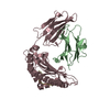
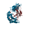
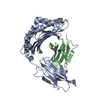
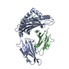
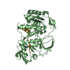
Entry Database : PDB / ID : 4wsiTitle Crystal Structure of PALS1/Crb complex MAGUK p55 subfamily member 5 Protein crumbs Keywords / Function / homology Function Domain/homology Component
/ / / / / / / / / / / / / / / / / / / / / / / / / / / / / / / / / / / / / / / / / / / / / / / / / / / / / / / / / / / / / / / / / / / / / / / / / / / / / / / / / / / / / / / / / / / / / / / / / / / / / / / / / / / / / / / / / / / / / / / / / / / / / / / / / / / / / / / / / / / / / / / / / / / / / / / / / / / / / / / / / / / Biological species Homo sapiens (human)Drosophila melanogaster (fruit fly)Method / / / Resolution : 2.95 Å Authors Wei, Z. / Li, Y. / Zhang, M. Journal : Proc.Natl.Acad.Sci.USA / Year : 2014Title : Structure of Crumbs tail in complex with the PALS1 PDZ-SH3-GK tandem reveals a highly specific assembly mechanism for the apical Crumbs complex.Authors : Li, Y. / Wei, Z. / Yan, Y. / Wan, Q. / Du, Q. / Zhang, M. History Deposition Oct 28, 2014 Deposition site / Processing site Revision 1.0 Nov 26, 2014 Provider / Type Revision 1.1 Feb 11, 2015 Group Revision 1.2 Mar 20, 2024 Group Data collection / Database references ... Data collection / Database references / Derived calculations / Source and taxonomy Category chem_comp_atom / chem_comp_bond ... chem_comp_atom / chem_comp_bond / citation / database_2 / entity_src_gen / pdbx_struct_oper_list Item _citation.journal_id_CSD / _database_2.pdbx_DOI ... _citation.journal_id_CSD / _database_2.pdbx_DOI / _database_2.pdbx_database_accession / _entity_src_gen.pdbx_alt_source_flag / _pdbx_struct_oper_list.symmetry_operation
Show all Show less
 Movie
Movie Controller


 Open data
 Basic information
 Components
 Keywords
 Function and homology information
 Homo sapiens (human)
 X-RAY DIFFRACTION /
  Authors
 Citation
 Structure visualization
 Downloads & links
 Other downloads
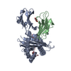


 PDBj
PDBj


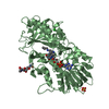
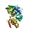
 Assembly
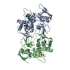


 Mass: 18.015 Da / Num. of mol.: 20 / Source method: isolated from a natural source / Formula: H2O
 Sample preparation
 / Beamline: BL17U / Wavelength: 0.98 Å
 Processing