 Movie
Movie Controller

[English] 日本語
 Yorodumi
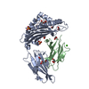
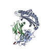
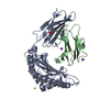
- PDB-1ldp: CRYSTAL STRUCTURE OF MURINE MHC CLASS I H-2LD WITH A MIXTURE OF B... -
+ Open data

- Basic information
| Entry | Database: PDB / ID: 1ldp | |||||||||
|---|---|---|---|---|---|---|---|---|---|---|
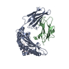
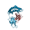
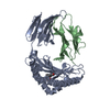
| Title | CRYSTAL STRUCTURE OF MURINE MHC CLASS I H-2LD WITH A MIXTURE OF BOUND PEPTIDES | |||||||||
 Components Components |
| |||||||||
 Keywords Keywords | COMPLEX (MHC I/PEPTIDE) / COMPLEX (MHC I-PEPTIDE) / MAJOR HISTOCOMPATIBILITY COMPLEX / IMMUNOLOGY / CELLULAR IMMUNITY / PEPTIDE ANTIGEN / CELL SURFACE RECEPTOR / ANTIGEN RECEPTOR / ANTIGEN PRESENTATION / COMPLEX (MHC I-PEPTIDE) complex | |||||||||
| Function / homology |  Function and homology information Function and homology informationMHC class Ib protein complex / natural killer cell lectin-like receptor binding / TAP2 binding / TAP1 binding / cis-Golgi network membrane / Endosomal/Vacuolar pathway / DAP12 interactions / Antigen Presentation: Folding, assembly and peptide loading of class I MHC / ER-Phagosome pathway / DAP12 signaling ...MHC class Ib protein complex / natural killer cell lectin-like receptor binding / TAP2 binding / TAP1 binding / cis-Golgi network membrane / Endosomal/Vacuolar pathway / DAP12 interactions / Antigen Presentation: Folding, assembly and peptide loading of class I MHC / ER-Phagosome pathway / DAP12 signaling / Immunoregulatory interactions between a Lymphoid and a non-Lymphoid cell / TAP complex binding / Golgi medial cisterna / regulation of membrane depolarization / CD8 receptor binding / beta-2-microglobulin binding / endoplasmic reticulum exit site / TAP binding / MHC class I protein binding / antigen processing and presentation of endogenous peptide antigen via MHC class Ib / antigen processing and presentation of endogenous peptide antigen via MHC class I via ER pathway, TAP-independent / cellular defense response / T cell receptor binding / Neutrophil degranulation / 14-3-3 protein binding / lumenal side of endoplasmic reticulum membrane / regulation of iron ion transport / cellular response to iron(III) ion / negative regulation of iron ion transport / negative regulation of forebrain neuron differentiation / antigen processing and presentation of exogenous protein antigen via MHC class Ib, TAP-dependent / iron ion transport / peptide antigen assembly with MHC class I protein complex / regulation of erythrocyte differentiation / response to molecule of bacterial origin / defense response / HFE-transferrin receptor complex / MHC class I peptide loading complex / transferrin transport / cellular response to iron ion / negative regulation of receptor-mediated endocytosis / positive regulation of T cell cytokine production / antigen processing and presentation of endogenous peptide antigen via MHC class I / MHC class I protein complex / peptide antigen assembly with MHC class II protein complex / negative regulation of neurogenesis / MHC class II protein complex / cellular response to nicotine / positive regulation of receptor-mediated endocytosis / multicellular organismal-level iron ion homeostasis / positive regulation of T cell mediated cytotoxicity / antigen processing and presentation of exogenous peptide antigen via MHC class II / positive regulation of immune response / peptide antigen binding / phagocytic vesicle membrane / positive regulation of T cell activation / negative regulation of epithelial cell proliferation / sensory perception of smell / positive regulation of cellular senescence / MHC class II protein complex binding / T cell differentiation in thymus / late endosome membrane / antimicrobial humoral immune response mediated by antimicrobial peptide / negative regulation of neuron projection development / antibacterial humoral response / protein refolding / protein-folding chaperone binding / cellular response to lipopolysaccharide / early endosome membrane / amyloid fibril formation / protein homotetramerization / defense response to Gram-negative bacterium / intracellular iron ion homeostasis / learning or memory / early endosome / defense response to Gram-positive bacterium / immune response / receptor ligand activity / signaling receptor binding / Golgi membrane / external side of plasma membrane / innate immune response / lysosomal membrane / structural molecule activity / cell surface / Golgi apparatus / endoplasmic reticulum / protein homodimerization activity / : / identical protein binding / plasma membrane / cytosol Similarity search - Function | |||||||||
| Biological species |   | |||||||||
| Method |  X-RAY DIFFRACTION / MOLECULAR REPLACEMENT / Resolution: 3.1 Å X-RAY DIFFRACTION / MOLECULAR REPLACEMENT / Resolution: 3.1 Å | |||||||||
 Authors Authors | Speir, J.A. / Wilson, I.A. | |||||||||
 Citation Citation | Journal: Immunity / Year: 1998 Title: Structural basis of 2C TCR allorecognition of H-2Ld peptide complexes. Authors: Speir, J.A. / Garcia, K.C. / Brunmark, A. / Degano, M. / Peterson, P.A. / Teyton, L. / Wilson, I.A. #1: Journal: Science / Year: 1998Title: Structural Basis of Plasticity in T Cell Receptor Recognition of a Self Peptide-Mhc Antigen Authors: Garcia, K.C. / Degano, M. / Pease, L.R. / Huang, M. / Peterson, P.A. / Teyton, L. / Wilson, I.A. | |||||||||
| History |
|
- Structure visualization
| Structure viewer | Molecule: MolmilJmol/JSmol |
|---|
- Downloads & links
-Download
| PDBx/mmCIF format | 1ldp.cif.gz | 87.8 KB | Display | PDBx/mmCIF format |
|---|---|---|---|---|
| PDB format | pdb1ldp.ent.gz | 62.8 KB | Display | PDB format |
| PDBx/mmJSON format | 1ldp.json.gz | Tree view | PDBx/mmJSON format | |
| Others |  Other downloads Other downloads |
-Validation report
| Arichive directory | https://data.pdbj.org/pub/pdb/validation_reports/ld/1ldpftp://data.pdbj.org/pub/pdb/validation_reports/ld/1ldp | HTTPS FTP |
|---|
-Related structure data
| Related structure data | 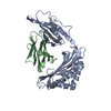 1hocS S: Starting model for refinement |
|---|---|
| Similar structure data |


-Links
 PDBj
PDBj


- Assembly
| Deposited unit | 
| ||||||||
|---|---|---|---|---|---|---|---|---|---|
| 1 |
| ||||||||
| Unit cell |
|
-Components
-MHC CLASS I H- ... , 2 types, 2 molecules HL
| #1: Protein | Mass: 31388.924 Da / Num. of mol.: 1 Fragment: CHAIN H IS THE HEAVY CHAIN, PEPTIDE BINDING DOMAIN, CHAIN L IS THE LIGHT CHAIN OR BETA-2-MICROGLOBULIN Mutation: HEAVY CHAIN TRUNCATED AFTER RESIDUE 274, B2M, LIGHT CHAIN TRUNCATED AFTER RESIDUE 99 Source method: isolated from a genetically manipulated source Details: ASPARAGINE LINKED N-ACETYL-GLUCOSAMINE CARBOHYDRATES AT CHAIN H, N86 AND N176 Source: (gene. exp.) |
|---|---|
| #2: Protein | Mass: 11704.359 Da / Num. of mol.: 1 Fragment: CHAIN H IS THE HEAVY CHAIN, PEPTIDE BINDING DOMAIN, CHAIN L IS THE LIGHT CHAIN OR BETA-2-MICROGLOBULIN Mutation: HEAVY CHAIN TRUNCATED AFTER RESIDUE 274, B2M, LIGHT CHAIN TRUNCATED AFTER RESIDUE 99 Source method: isolated from a genetically manipulated source Details: ASPARAGINE LINKED N-ACETYL-GLUCOSAMINE CARBOHYDRATES AT CHAIN H, N86 AND N176 Source: (gene. exp.) |
-Protein/peptide , 2 types, 2 molecules PQ
| #3: Protein/peptide | Mass: 743.870 Da / Num. of mol.: 1 Fragment: CHAIN P IS A PEPTIDE, CHAIN Q IS A MODEL OF PEPTIDE QL9 DERIVED FROM P Source method: isolated from a natural source Details: CHAIN P PEPTIDE IS THE NATURAL PRODUCT OF THE EXPRESSION SYSTEM. IT IS ACTUALLY A MODEL REPRESENTING THE ELECTRON DENSITY OF A MIXTURE OF BOUND PEPTIDES AVAILABLE IN THE PROTEIN EXPRESSION MEDIA. Source: (natural) |
|---|---|
| #4: Protein/peptide | Mass: 1063.202 Da / Num. of mol.: 1 Fragment: CHAIN P IS A PEPTIDE, CHAIN Q IS A MODEL OF PEPTIDE QL9 DERIVED FROM P Source method: isolated from a natural source Details: CHAIN P PEPTIDE IS THE NATURAL PRODUCT OF THE EXPRESSION SYSTEM. IT IS ACTUALLY A MODEL REPRESENTING THE ELECTRON DENSITY OF A MIXTURE OF BOUND PEPTIDES AVAILABLE IN THE PROTEIN EXPRESSION MEDIA. Source: (natural) |
-Sugars , 2 types, 2 molecules 
| #5: Polysaccharide | 2-acetamido-2-deoxy-alpha-D-glucopyranose-(1-4)-2-acetamido-2-deoxy-beta-D-glucopyranose Source method: isolated from a genetically manipulated source |
|---|---|
| #6: Sugar | ChemComp-NAG /  Type: D-saccharide, beta linking / Mass: 221.208 Da / Num. of mol.: 1 Type: D-saccharide, beta linking / Mass: 221.208 Da / Num. of mol.: 1Source method: isolated from a genetically manipulated source Formula: C8H15NO6 |
-Details
| Has protein modification | Y |
|---|
-Experimental details
-Experiment
| Experiment | Method: X-RAY DIFFRACTION / Number of used crystals: 3 |
|---|
- Sample preparation
| Crystal | Density Matthews: 3.18 Å3/Da / Density % sol: 61 % | ||||||||||||||||||||||||||||||||||||
|---|---|---|---|---|---|---|---|---|---|---|---|---|---|---|---|---|---|---|---|---|---|---|---|---|---|---|---|---|---|---|---|---|---|---|---|---|---|
| Crystal grow | pH: 6.5 Details: PROTEIN WAS CRYSTALLIZED FROM 100MM SODIUM CACODYLATE, 0.2M AMMONIUM SULFATE, 0.5M SODIUM BROMIDE, 30% PEG 8000, PH 6.5. | ||||||||||||||||||||||||||||||||||||
| Crystal grow | *PLUS Method: unknown | ||||||||||||||||||||||||||||||||||||
| Components of the solutions | *PLUS
|
-Data collection
| Diffraction | Mean temperature: 298 K |
|---|---|
| Diffraction source | Source: ROTATING ANODE / Type: ELLIOTT GX-18 / Wavelength: 1.5418 |
| Detector | Type: SIEMENS / Detector: AREA DETECTOR / Date: Jan 1, 1996 / Details: DOUBLE-MIRROR FOCUSING CAMERAS |
| Radiation | Monochromator: NI FILTER / Monochromatic (M) / Laue (L): M / Scattering type: x-ray |
| Radiation wavelength | Wavelength: 1.5418 Å / Relative weight: 1 |
| Reflection | Resolution: 2.8→15 Å / Num. obs: 11809 / % possible obs: 81 % / Observed criterion σ(I): 0 / Redundancy: 2.2 % / Rmerge(I) obs: 0.113 / Rsym value: 0.113 / Net I/σ(I): 5.6 |
| Reflection shell | Resolution: 2.8→2.9 Å / Redundancy: 1.4 % / Rmerge(I) obs: 0.358 / Mean I/σ(I) obs: 1.8 / Rsym value: 0.358 / % possible all: 63.6 |
| Reflection shell | *PLUS % possible obs: 63.6 % / Num. unique obs: 926 |
- Processing
| Software |
| ||||||||||||||||||||||||||||||||||||||||||||||||||||||||||||
|---|---|---|---|---|---|---|---|---|---|---|---|---|---|---|---|---|---|---|---|---|---|---|---|---|---|---|---|---|---|---|---|---|---|---|---|---|---|---|---|---|---|---|---|---|---|---|---|---|---|---|---|---|---|---|---|---|---|---|---|---|---|
| Refinement | Method to determine structure: MOLECULAR REPLACEMENT Starting model: PDB ENTRY 1HOC Resolution: 3.1→6 Å / Rfactor Rfree error: 0.016 / Data cutoff high absF: 100000 / Data cutoff low absF: 0.1 / Isotropic thermal model: ONE B-VALUE PER RESIDUE / Cross valid method: THROUGHOUT / σ(F): 1 Details: SOME LIBRARIES WERE MODIFIED IN-HOUSE TO PROVIDE MISSING CARBOHYDRATE AND PROTEIN SPECIFICATIONS. X-PLOR 3.1 ALSO WAS USED.
| ||||||||||||||||||||||||||||||||||||||||||||||||||||||||||||
| Displacement parameters | Biso mean: 28 Å2
| ||||||||||||||||||||||||||||||||||||||||||||||||||||||||||||
| Refine analyze |
| ||||||||||||||||||||||||||||||||||||||||||||||||||||||||||||
| Refinement step | Cycle: LAST / Resolution: 3.1→6 Å
| ||||||||||||||||||||||||||||||||||||||||||||||||||||||||||||
| Refine LS restraints |
| ||||||||||||||||||||||||||||||||||||||||||||||||||||||||||||
| LS refinement shell | Resolution: 3.1→3.22 Å / Rfactor Rfree error: 0.062 / Total num. of bins used: 8
| ||||||||||||||||||||||||||||||||||||||||||||||||||||||||||||
| Xplor file |
| ||||||||||||||||||||||||||||||||||||||||||||||||||||||||||||
| Software | *PLUS Name: X-PLOR / Version: 3.825 / Classification: refinement | ||||||||||||||||||||||||||||||||||||||||||||||||||||||||||||
| Refinement | *PLUS Num. reflection obs: 7379 / Rfactor all: 0.222 | ||||||||||||||||||||||||||||||||||||||||||||||||||||||||||||
| Solvent computation | *PLUS | ||||||||||||||||||||||||||||||||||||||||||||||||||||||||||||
| Displacement parameters | *PLUS | ||||||||||||||||||||||||||||||||||||||||||||||||||||||||||||
| Refine LS restraints | *PLUS
| ||||||||||||||||||||||||||||||||||||||||||||||||||||||||||||
| LS refinement shell | *PLUS Rfactor obs: 0.291 |
