 Movie
Movie Controller

[English] 日本語
 Yorodumi
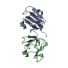
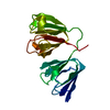
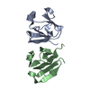
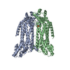
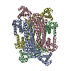
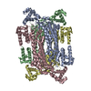
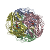
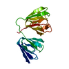
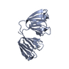
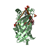
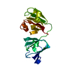
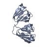
- PDB-1h4a: Human GammaD Crystallin R58H mutant structure AT 1.15 A resolution -
+ Open data

- Basic information
| Entry | Database: PDB / ID: 1h4a | ||||||
|---|---|---|---|---|---|---|---|
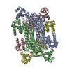
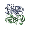
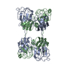
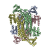
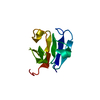
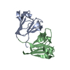
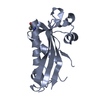
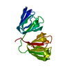
| Title | Human GammaD Crystallin R58H mutant structure AT 1.15 A resolution | ||||||
 Components Components | Gamma-crystallin D | ||||||
 Keywords Keywords | EYE LENS PROTEIN / CRYSTALLIN | ||||||
| Function / homology |  Function and homology information Function and homology informationlens fiber cell differentiation / structural constituent of eye lens / lens development in camera-type eye / visual perception / cellular response to reactive oxygen species / nucleus / cytoplasm Similarity search - Function | ||||||
| Biological species |  Homo sapiens (human) Homo sapiens (human) | ||||||
| Method |  X-RAY DIFFRACTION / SYNCHROTRON / MOLECULAR REPLACEMENT / Resolution: 1.15 Å X-RAY DIFFRACTION / SYNCHROTRON / MOLECULAR REPLACEMENT / Resolution: 1.15 Å | ||||||
 Authors Authors | Basak, A.K. / Slingsby, C. | ||||||
 Citation Citation | Journal: J.Mol.Biol. / Year: 2003 Title: High-Resolution X-Ray Crystal Structures of Human GammaD Crystallin (1.25A) and the R58H Mutant (1.15A) Associated with Aculeiform Cataract Authors: Basak, A.K. / Bateman, O. / Slingsby, C. / Pande, A. / Asherie, N. / Ogun, O. / Benedek, G. / Pande, J. #1: Journal: Exp.Eye Res. / Year: 1991 Title: Crystal Structure of Calf Eye Lens Gamma-Crystallin Iiib at 2.5 A Resolution: Its Relation to Function Authors: Yu, C. / Nevskaya, N. / Vernoslova, E. / Nikonov, S. / Yu, S. / Brazhnikov, E. / Fomenkova, N. / Lunin, V. / Urzhumtsev, A. #2: Journal: Proteins: Struct.,Funct., Genet. / Year: 1988 Title: Surface Interactions of Gamma-Crystallins in the Crystal Medium in Relation to Their Association in the Eye Lens Authors: Sergeev, Y.V. / Chirgadze, Y.N. / Mylvaganam, S.E. / Driessen, H. / Slingsby, C. / Blundell, T.L. #3: Journal: Mol.Biol.(Engl.Transl.) / Year: 1987 Title: Key Role of Residue 103 in Surface Interactions of Gamma-Crystallins Authors: Sergeev Yu, V. / Chirgadze Yu, N. / Driessen, H. / Slingsby, C. / Blundell, T.L. #4: Journal: Acta Crystallogr.,Sect.A / Year: 1985Title: Phase Improvement in Protein Crystallography Using a Mixed Electron Density Model Authors: Lunin Yu, V. / Urzhumtsev, A.G. / Vernoslova, E.A. / Chirgadze Yu, N. / Nevskaya, N.A. / Fomenkova, N.P. #5: Journal: J.Mol.Biol. / Year: 1977 Title: Crystallographic Study of Gamma-Crystallins from Calf Lens Authors: Chirgadze, Y.N. / Nikonov, S.V. / Garber, M.B. / Reshetnikova, L.S. | ||||||
| History |
|
- Structure visualization
| Structure viewer | Molecule: MolmilJmol/JSmol |
|---|
- Downloads & links
-Download
| PDBx/mmCIF format | 1h4a.cif.gz | 95.9 KB | Display | PDBx/mmCIF format |
|---|---|---|---|---|
| PDB format | pdb1h4a.ent.gz | 72.4 KB | Display | PDB format |
| PDBx/mmJSON format | 1h4a.json.gz | Tree view | PDBx/mmJSON format | |
| Others |  Other downloads Other downloads |
-Validation report
| Arichive directory | https://data.pdbj.org/pub/pdb/validation_reports/h4/1h4aftp://data.pdbj.org/pub/pdb/validation_reports/h4/1h4a | HTTPS FTP |
|---|
-Related structure data
| Related structure data | 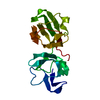 1hk0C 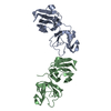 1elpS S: Starting model for refinement C: citing same article ( |
|---|---|
| Similar structure data |


-Links
 PDBj
PDBj
- Assembly
| Deposited unit | 
| ||||||||
|---|---|---|---|---|---|---|---|---|---|
| 1 |
| ||||||||
| Unit cell |
|
-Components
| #1: Protein | Mass: 20615.922 Da / Num. of mol.: 1 / Mutation: R58H Source method: isolated from a genetically manipulated source Source: (gene. exp.) Homo sapiens (human) / Gene: CRYGD, CRYG4 / Production host:  |
|---|---|
| #2: Water | ChemComp-HOH /  Mass: 18.015 Da / Num. of mol.: 276 / Source method: isolated from a natural source / Formula: H2O Mass: 18.015 Da / Num. of mol.: 276 / Source method: isolated from a natural source / Formula: H2O |
| Compound details | DOMINANT STRUCTURAL COMPONENTS OF THE VERTEBRATE EYE LENS. THESE PROTEINS HAVE A TWO-DOMAIN BETA- ...DOMINANT STRUCTURAL |
-Experimental details
-Experiment
| Experiment | Method: X-RAY DIFFRACTION / Number of used crystals: 1 |
|---|
- Sample preparation
| Crystal | Density Matthews: 1.94 Å3/Da / Density % sol: 39 % | ||||||||||||||||||
|---|---|---|---|---|---|---|---|---|---|---|---|---|---|---|---|---|---|---|---|
| Crystal grow | Temperature: 277 K / pH: 7 / Details: 10MM PHOSPHATE, 4 DEGREE CELSIUS, pH 7.00 | ||||||||||||||||||
| Crystal grow | *PLUS pH: 7 / Method: batch method | ||||||||||||||||||
| Components of the solutions | *PLUS
|
-Data collection
| Diffraction | Mean temperature: 100 K |
|---|---|
| Diffraction source | Source: SYNCHROTRON / Site: ESRF  / Beamline: ID14-1 / Wavelength: 0.934 / Beamline: ID14-1 / Wavelength: 0.934 |
| Detector | Type: MARRESEARCH / Detector: CCD / Date: Sep 15, 2001 / Details: MIRROR |
| Radiation | Monochromator: LAUE DIAMOND C111 / Protocol: SINGLE WAVELENGTH / Monochromatic (M) / Laue (L): M / Scattering type: x-ray |
| Radiation wavelength | Wavelength: 0.934 Å / Relative weight: 1 |
| Reflection | Resolution: 1.15→28.33 Å / Num. obs: 55087 / % possible obs: 95.3 % / Redundancy: 4.6 % / Rmerge(I) obs: 0.05 / Net I/σ(I): 9.2 |
| Reflection shell | Resolution: 1.15→1.21 Å / Redundancy: 2.7 % / Rmerge(I) obs: 0.109 / Mean I/σ(I) obs: 6.3 / % possible all: 97.6 |
| Reflection | *PLUS Num. measured all: 463412 / Rmerge(I) obs: 0.05 |
| Reflection shell | *PLUS % possible obs: 97.6 % |
- Processing
| Software |
| ||||||||||||||||||||
|---|---|---|---|---|---|---|---|---|---|---|---|---|---|---|---|---|---|---|---|---|---|
| Refinement | Method to determine structure: MOLECULAR REPLACEMENT Starting model: PDB ENTRY 1ELP Resolution: 1.15→27.02 Å / SU B: 0.504 / SU ML: 0.024 / Cross valid method: THROUGHOUT / σ(F): 0 / ESU R: 0.042 / ESU R Free: 0.041 / Details: HYDROGENS HAVE BEEN ADDED IN THE RIDING POSITIONS
| ||||||||||||||||||||
| Displacement parameters | Biso mean: 8.566 Å2
| ||||||||||||||||||||
| Refinement step | Cycle: LAST / Resolution: 1.15→27.02 Å
| ||||||||||||||||||||
| Refinement | *PLUS Lowest resolution: 28.33 Å / % reflection Rfree: 8 % / Rfactor obs: 0.1576 / Rfactor Rfree: 0.1822 / Rfactor Rwork: 0.1555 | ||||||||||||||||||||
| Solvent computation | *PLUS | ||||||||||||||||||||
| Displacement parameters | *PLUS | ||||||||||||||||||||
| Refine LS restraints | *PLUS
|
