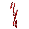
 Movie
Movie Controller


+ Open data

- Basic information
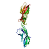
| Entry | Database: PDB / ID: 1a45 | |||||||||
|---|---|---|---|---|---|---|---|---|---|---|
| Title | GAMMAF CRYSTALLIN FROM BOVINE LENS | |||||||||
 Components Components | GAMMAF CRYSTALLIN | |||||||||
 Keywords Keywords | EYE LENS PROTEIN | |||||||||
| Function / homology |  Function and homology information Function and homology informationstructural constituent of eye lens / lens development in camera-type eye / visual perception Similarity search - Function | |||||||||
| Biological species |  | |||||||||
| Method |  X-RAY DIFFRACTION / SYNCHROTRON / MOLECULAR REPLACEMENT / Resolution: 2.3 Å X-RAY DIFFRACTION / SYNCHROTRON / MOLECULAR REPLACEMENT / Resolution: 2.3 Å | |||||||||
 Authors Authors | Norledge, B.V. / Hay, R. / Bateman, O.A. / Slingsby, C. / White, H.E. / Moss, D.S. / Lindley, P.F. / Driessen, H.P.C. | |||||||||
 Citation Citation | Journal: Exp.Eye Res. / Year: 1997 Title: Towards a molecular understanding of phase separation in the lens: a comparison of the X-ray structures of two high Tc gamma-crystallins, gammaE and gammaF, with two low Tc gamma-crystallins, gammaB and gammaD. Authors: Norledge, B.V. / Hay, R.E. / Bateman, O.A. / Slingsby, C. / Driessen, H.P. #1: Journal: J.Mol.Biol. / Year: 1989Title: Packing Interactions in the Eye-Lens. Structural Analysis, Internal Symmetry and Lattice Interactions of Bovine Gamma Iva-Crystallin Authors: White, H.E. / Driessen, H.P. / Slingsby, C. / Moss, D.S. / Lindley, P.F. #2: Journal: Acta Crystallogr.,Sect.B / Year: 1988Title: The Use of Pseudosymmetry in the Rotation Function of Gamma Iva-Crystallin Authors: White, H.E. / Driessen, H.P. / Slingsby, C. / Moss, D.S. / Turnell, W.G. / Lindley, P.F. #3: Journal: Exp.Eye Res. / Year: 1983Title: Purification and Crystallization of Mammalian Lens Gamma-Crystallins Authors: Slingsby, C. / Miller, L.R. #4: Journal: Acta Crystallogr.,Sect.B / Year: 1978Title: The Low-Resolution Structure Analysis of the Lens Protein Gamma-Crystallin Authors: Blundell, T.L. / Lindley, P.F. / Moss, D.S. / Slingsby, C. / Tickle, I.J. / Turnell, W.G. | |||||||||
| History |
|
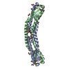
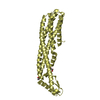
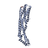
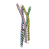
- Structure visualization
| Structure viewer | Molecule: MolmilJmol/JSmol |
|---|
- Downloads & links
-Download
| PDBx/mmCIF format | 1a45.cif.gz | 53.1 KB | Display | PDBx/mmCIF format |
|---|---|---|---|---|
| PDB format | pdb1a45.ent.gz | 38.1 KB | Display | PDB format |
| PDBx/mmJSON format | 1a45.json.gz | Tree view | PDBx/mmJSON format | |
| Others |  Other downloads Other downloads |
-Validation report
| Arichive directory | https://data.pdbj.org/pub/pdb/validation_reports/a4/1a45ftp://data.pdbj.org/pub/pdb/validation_reports/a4/1a45 | HTTPS FTP |
|---|
-Related structure data
| Related structure data |  1a5dC  4gcrS S: Starting model for refinement C: citing same article ( |
|---|---|
| Similar structure data |


-Links
 PDBj
PDBj
- Assembly
| Deposited unit | 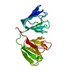
| ||||||||
|---|---|---|---|---|---|---|---|---|---|
| 1 | 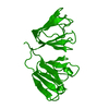
| ||||||||
| Unit cell |
|
-Components
| #1: Protein | Mass: 20985.273 Da / Num. of mol.: 1 / Source method: isolated from a natural source / Source: (natural) |
|---|---|
| #2: Water | ChemComp-HOH /  Mass: 18.015 Da / Num. of mol.: 207 / Source method: isolated from a natural source / Formula: H2O Mass: 18.015 Da / Num. of mol.: 207 / Source method: isolated from a natural source / Formula: H2O |
| Sequence details | THE RESIDUES HAVE BEEN NUMBERED ACCORDING TO THE HOMOLOGY WITH BOVINE LENS GAMMA B-CRYSTALLIN. ...THE RESIDUES HAVE BEEN NUMBERED ACCORDING TO THE HOMOLOGY WITH BOVINE LENS GAMMA B-CRYSTALLIN |
-Experimental details
-Experiment
| Experiment | Method: X-RAY DIFFRACTION / Number of used crystals: 3 |
|---|
- Sample preparation
| Crystal | Density Matthews: 1.8 Å3/Da / Density % sol: 32 % Description: DATA WERE COLLECTED USING THE OSCILLATION METHOD ON AN ARNDT-WONACOTT CAMERA. THERE IS INCOMPLETE DATA TO 2.1 A. | |||||||||||||||
|---|---|---|---|---|---|---|---|---|---|---|---|---|---|---|---|---|
| Crystal grow | pH: 7 / Details: pH 7.0 | |||||||||||||||
| Crystal grow | *PLUS pH: 6.86 / Method: vapor diffusion, hanging drop | |||||||||||||||
| Components of the solutions | *PLUS
|
-Data collection
| Diffraction | Mean temperature: 300 K |
|---|---|
| Diffraction source | Source: SYNCHROTRON / Site: SRS  / Beamline: PX7.2 / Wavelength: 1.5418 / Beamline: PX7.2 / Wavelength: 1.5418 |
| Detector | Detector: FILM / Date: Jun 1, 1983 |
| Radiation | Monochromator: GRAPHITE(002) / Monochromatic (M) / Laue (L): M / Scattering type: x-ray |
| Radiation wavelength | Wavelength: 1.5418 Å / Relative weight: 1 |
| Reflection | Resolution: 2.3→24 Å / Num. obs: 6300 / % possible obs: 88.7 % / Redundancy: 4 % / Rmerge(I) obs: 0.104 |
- Processing
| Software |
| ||||||||||||||||||||||||||||||||||||||||||||||||||||||||||||
|---|---|---|---|---|---|---|---|---|---|---|---|---|---|---|---|---|---|---|---|---|---|---|---|---|---|---|---|---|---|---|---|---|---|---|---|---|---|---|---|---|---|---|---|---|---|---|---|---|---|---|---|---|---|---|---|---|---|---|---|---|---|
| Refinement | Method to determine structure: MOLECULAR REPLACEMENT Starting model: PDB ENTRY 4GCR Resolution: 2.3→8 Å / σ(F): 0 Details: THE AUTHORS ALSO USED REFINEMENT PROGRAM RESTRAIN (DRIESSEN, HANEEF, HARRIS, HOWLIN, KHAN & MOSS, J. APPL. CRYSTALLOGR. 22, 510, 1989).
| ||||||||||||||||||||||||||||||||||||||||||||||||||||||||||||
| Refinement step | Cycle: LAST / Resolution: 2.3→8 Å
| ||||||||||||||||||||||||||||||||||||||||||||||||||||||||||||
| Refine LS restraints |
| ||||||||||||||||||||||||||||||||||||||||||||||||||||||||||||
| Software | *PLUS Name: X-PLOR / Version: 3.1 / Classification: refinement | ||||||||||||||||||||||||||||||||||||||||||||||||||||||||||||
| Refine LS restraints | *PLUS
|
