 Movie
Movie Controller


+ Open data

- Basic information
| Entry | Database: PDB / ID: 1a5d | ||||||
|---|---|---|---|---|---|---|---|
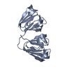
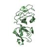
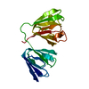
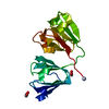
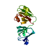
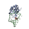
| Title | GAMMAE CRYSTALLIN FROM RAT LENS | ||||||
 Components Components | GAMMAE CRYSTALLIN | ||||||
 Keywords Keywords | EYE LENS PROTEIN | ||||||
| Function / homology |  Function and homology information Function and homology informationstructural constituent of eye lens / lens development in camera-type eye / visual perception Similarity search - Function | ||||||
| Biological species |  | ||||||
| Method |  X-RAY DIFFRACTION / MOLECULAR REPLACEMENT / Resolution: 2.3 Å X-RAY DIFFRACTION / MOLECULAR REPLACEMENT / Resolution: 2.3 Å | ||||||
 Authors Authors | Norledge, B.V. / Hay, R. / Bateman, O.A. / Slingsby, C. / Driessen, H.P.C. | ||||||
 Citation Citation | Journal: Exp.Eye Res. / Year: 1997 Title: Towards a molecular understanding of phase separation in the lens: a comparison of the X-ray structures of two high Tc gamma-crystallins, gammaE and gammaF, with two low Tc gamma-crystallins, gammaB and gammaD. Authors: Norledge, B.V. / Hay, R.E. / Bateman, O.A. / Slingsby, C. / Driessen, H.P. | ||||||
| History |
|
- Structure visualization
| Structure viewer | Molecule: MolmilJmol/JSmol |
|---|
- Downloads & links
-Download
| PDBx/mmCIF format | 1a5d.cif.gz | 88.5 KB | Display | PDBx/mmCIF format |
|---|---|---|---|---|
| PDB format | pdb1a5d.ent.gz | 67.9 KB | Display | PDB format |
| PDBx/mmJSON format | 1a5d.json.gz | Tree view | PDBx/mmJSON format | |
| Others |  Other downloads Other downloads |
-Validation report
| Arichive directory | https://data.pdbj.org/pub/pdb/validation_reports/a5/1a5dftp://data.pdbj.org/pub/pdb/validation_reports/a5/1a5d | HTTPS FTP |
|---|
-Related structure data
| Related structure data |  1a45C  2gcr S: Starting model for refinement C: citing same article ( |
|---|---|
| Similar structure data |


-Links
 PDBj
PDBj
- Assembly
| Deposited unit | 
| ||||||||
|---|---|---|---|---|---|---|---|---|---|
| 1 | 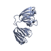
| ||||||||
| 2 | 
| ||||||||
| Unit cell |
| ||||||||
| Noncrystallographic symmetry (NCS) | NCS oper: (Code: given Matrix: (0.9997, 0.0217, -0.0055), Vector: Details | THE MOST LIKELY ACTIVE BIOLOGICAL STATE IS MONOMERIC. | |
-Components
| #1: Protein | Mass: 21163.611 Da / Num. of mol.: 2 / Source method: isolated from a natural source / Source: (natural) #2: Water | ChemComp-HOH / |  Mass: 18.015 Da / Num. of mol.: 257 / Source method: isolated from a natural source / Formula: H2O Mass: 18.015 Da / Num. of mol.: 257 / Source method: isolated from a natural source / Formula: H2O |
|---|
-Experimental details
-Experiment
| Experiment | Method: X-RAY DIFFRACTION / Number of used crystals: 1 |
|---|
- Sample preparation
| Crystal | Density Matthews: 1.99 Å3/Da / Density % sol: 38.05 % | ||||||||||||||||||||||||||||||||||||||||||
|---|---|---|---|---|---|---|---|---|---|---|---|---|---|---|---|---|---|---|---|---|---|---|---|---|---|---|---|---|---|---|---|---|---|---|---|---|---|---|---|---|---|---|---|
| Crystal grow | pH: 7 / Details: pH 7.0 | ||||||||||||||||||||||||||||||||||||||||||
| Crystal grow | *PLUS pH: 6.86 / Method: vapor diffusion, hanging drop | ||||||||||||||||||||||||||||||||||||||||||
| Components of the solutions | *PLUS
|
-Data collection
| Diffraction | Mean temperature: 300 K |
|---|---|
| Diffraction source | Type: BRUKER NONIUS / Wavelength: 1.5418 |
| Detector | Type: ENRAF-NONIUS FAST / Detector: DIFFRACTOMETER / Date: Jun 1, 1989 |
| Radiation | Monochromatic (M) / Laue (L): M / Scattering type: x-ray |
| Radiation wavelength | Wavelength: 1.5418 Å / Relative weight: 1 |
| Reflection | Resolution: 2.3→50.9 Å / Num. obs: 12942 / % possible obs: 84.7 % / Redundancy: 3.2 % / Rmerge(I) obs: 0.082 |
| Reflection | *PLUS Num. measured all: 41615 |
- Processing
| Software |
| ||||||||||||||||||||||||||||||||||||||||||||||||||||||||||||
|---|---|---|---|---|---|---|---|---|---|---|---|---|---|---|---|---|---|---|---|---|---|---|---|---|---|---|---|---|---|---|---|---|---|---|---|---|---|---|---|---|---|---|---|---|---|---|---|---|---|---|---|---|---|---|---|---|---|---|---|---|---|
| Refinement | Method to determine structure: MOLECULAR REPLACEMENT Starting model: PDB ENTRY 2GCR 2gcr Resolution: 2.3→8 Å / σ(F): 0 Details: THE AUTHORS ALSO USED REFINEMENT PROGRAM RESTRAIN (DRIESSEN, HANEEF, HARRIS, HOWLIN, KHAN & MOSS, J. APPL. CRYSTALLOGR. 22, 510, 1989). THERE WAS NO ELECTRON DENSITY FOR THE SIDE CHAINS OF ...Details: THE AUTHORS ALSO USED REFINEMENT PROGRAM RESTRAIN (DRIESSEN, HANEEF, HARRIS, HOWLIN, KHAN & MOSS, J. APPL. CRYSTALLOGR. 22, 510, 1989). THERE WAS NO ELECTRON DENSITY FOR THE SIDE CHAINS OF SER B 58 AND SER B 119.
| ||||||||||||||||||||||||||||||||||||||||||||||||||||||||||||
| Refinement step | Cycle: LAST / Resolution: 2.3→8 Å
| ||||||||||||||||||||||||||||||||||||||||||||||||||||||||||||
| Refine LS restraints |
| ||||||||||||||||||||||||||||||||||||||||||||||||||||||||||||
| Software | *PLUS Name: X-PLOR / Version: 3.1 / Classification: refinement | ||||||||||||||||||||||||||||||||||||||||||||||||||||||||||||
| Refine LS restraints | *PLUS
|
