 Movie
Movie Controller


+ Open data

- Basic information
| Entry | Database: PDB / ID: 1i5i | ||||||
|---|---|---|---|---|---|---|---|
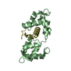
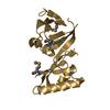
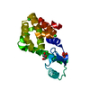
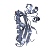
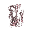
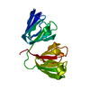
| Title | THE C18S MUTANT OF BOVINE (GAMMA-B)-CRYSTALLIN | ||||||
 Components Components | (GAMMA-B) CRYSTALLIN | ||||||
 Keywords Keywords | STRUCTURAL PROTEIN / EYE LENS PROTEIN / CRYSTALLIN | ||||||
| Function / homology |  Function and homology information Function and homology informationstructural constituent of eye lens / lens development in camera-type eye / visual perception Similarity search - Function | ||||||
| Biological species |  | ||||||
| Method |  X-RAY DIFFRACTION / MOLECULAR REPLACEMENT / Resolution: 2.4 Å X-RAY DIFFRACTION / MOLECULAR REPLACEMENT / Resolution: 2.4 Å | ||||||
 Authors Authors | Zarutskie, J.A. / Asherie, N. / Pande, J. / Pande, A. / Lomakin, J. / Lomakin, A. / Ogun, O. / Stern, L.J. / King, J.A. / Benedek, G.B. | ||||||
 Citation Citation | Journal: J.Mol.Biol. / Year: 2001 Title: Enhanced crystallization of the Cys18 to Ser mutant of bovine gammaB crystallin. Authors: Asherie, N. / Pande, J. / Pande, A. / Zarutskie, J.A. / Lomakin, J. / Lomakin, A. / Ogun, O. / Stern, L.J. / King, J. / Benedek, G.B. #1: Journal: Acta Crystallogr.,Sect.D / Year: 1993Title: Structure of the Bovine Eye Lens Protein Gamma-B (Gamma-II)-Crystallin at 1.47 Angstroms Authors: Najmudin, S. / Nalini, V. / Driessen, H.P.C. / Slingsby, C. / Blundell, T.L. / Moss, D.S. / Lindley, P.F. #2: Journal: Pept.Protein Rev. / Year: 1984Title: X-Ray Studies of the Lens Specific Proteins, the Crystallins Authors: Summers, L. / Wistow, G. / Narebor, M. / Moss, D. / Lindley, P. / Slingsby, C. / Blundell, T. / Bartunik, H. / Bartels, K. #3: Journal: J.Mol.Biol. / Year: 1983Title: X-ray analysis of the eye lens protein Gamma-II Crystallin at 1.9 Angstrom Resolution Authors: Wistow, G. / Turnell, B. / Summers, L. / Slingsby, C. / Moss, D. / Miller, L. / Lindley, P. / Blundell, T. #4: Journal: Nature / Year: 1981Title: The molecular structure and stability of the eye lens: X-Ray Analysis of Gamma-Crystallin II Authors: Blundell, T. / Lindley, P. / Miller, L. / Moss, D. / Slingsby, C. / Tickle, I. / Turnell, B. / Wistow, G. | ||||||
| History |
|
- Structure visualization
| Structure viewer | Molecule: MolmilJmol/JSmol |
|---|
- Downloads & links
-Download
| PDBx/mmCIF format | 1i5i.cif.gz | 50.1 KB | Display | PDBx/mmCIF format |
|---|---|---|---|---|
| PDB format | pdb1i5i.ent.gz | 35.2 KB | Display | PDB format |
| PDBx/mmJSON format | 1i5i.json.gz | Tree view | PDBx/mmJSON format | |
| Others |  Other downloads Other downloads |
-Validation report
| Arichive directory | https://data.pdbj.org/pub/pdb/validation_reports/i5/1i5iftp://data.pdbj.org/pub/pdb/validation_reports/i5/1i5i | HTTPS FTP |
|---|
-Related structure data
| Related structure data |  4gcrS S: Starting model for refinement |
|---|---|
| Similar structure data |


-Links
 PDBj
PDBj
- Assembly
| Deposited unit | 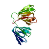
| ||||||||
|---|---|---|---|---|---|---|---|---|---|
| 1 |
| ||||||||
| Unit cell |
|
-Components
| #1: Protein | ( Mass: 20976.494 Da / Num. of mol.: 1 / Mutation: C18S Source method: isolated from a genetically manipulated source Source: (gene. exp.)  |
|---|---|
| #2: Water | ChemComp-HOH /  Mass: 18.015 Da / Num. of mol.: 71 / Source method: isolated from a natural source / Formula: H2O Mass: 18.015 Da / Num. of mol.: 71 / Source method: isolated from a natural source / Formula: H2O |
-Experimental details
-Experiment
| Experiment | Method: X-RAY DIFFRACTION / Number of used crystals: 1 |
|---|
- Sample preparation
| Crystal | Density Matthews: 1.97 Å3/Da / Density % sol: 37.49 % |
|---|---|
| Crystal grow | Temperature: 295 K Method: chill tube containing protein to 265 k, then bring to 295 k pH: 7 Details: 100 mM Phosphate, 20 mM DTT, pH 7.0, chill tube containing protein to 265 K, then bring to 295 K |
-Data collection
| Diffraction | Mean temperature: 296 K |
|---|---|
| Diffraction source | Source: ROTATING ANODE / Type: RIGAKU RU200 / Wavelength: 1.5418 |
| Detector | Type: RIGAKU RAXIS II / Detector: IMAGE PLATE / Date: Jun 26, 1999 / Details: MIRRORS |
| Radiation | Monochromator: Ni Filter / Protocol: SINGLE WAVELENGTH / Monochromatic (M) / Laue (L): M / Scattering type: x-ray |
| Radiation wavelength | Wavelength: 1.5418 Å / Relative weight: 1 |
| Reflection | Resolution: 2.4→20 Å / Num. obs: 6843 / % possible obs: 97.1 % / Observed criterion σ(I): 2 / Redundancy: 10 % / Biso Wilson estimate: 28 Å2 / Rmerge(I) obs: 0.074 / Rsym value: 0.074 / Net I/σ(I): 21.6 |
| Reflection shell | Resolution: 2.4→2.44 Å / Redundancy: 7.5 % / Rmerge(I) obs: 0.146 / Mean I/σ(I) obs: 9.6 / Num. unique all: 6843 / Rsym value: 0.146 / % possible all: 92.8 |
- Processing
| Software |
| ||||||||||||||||||||
|---|---|---|---|---|---|---|---|---|---|---|---|---|---|---|---|---|---|---|---|---|---|
| Refinement | Method to determine structure: MOLECULAR REPLACEMENT Starting model: 4GCR Resolution: 2.4→20 Å / Rfactor Rfree error: 0.009 / Data cutoff high absF: 100000 / Data cutoff low absF: 0.1 / Isotropic thermal model: RESTRAINED / Cross valid method: THROUGHOUT / σ(F): 2 / σ(I): 2 / Stereochemistry target values: Engh & Huber Details: Due to weak electron density in the region of the Tyrosine 174, a good model for the residue could not be refined, and is thus not included in the structure. Density was weak for the CG, SD, ...Details: Due to weak electron density in the region of the Tyrosine 174, a good model for the residue could not be refined, and is thus not included in the structure. Density was weak for the CG, SD, and CE of MET 160 and was not included in the structure.
| ||||||||||||||||||||
| Displacement parameters | Biso mean: 13.2 Å2
| ||||||||||||||||||||
| Refine analyze |
| ||||||||||||||||||||
| Refinement step | Cycle: LAST / Resolution: 2.4→20 Å
| ||||||||||||||||||||
| Refine LS restraints |
| ||||||||||||||||||||
| LS refinement shell | Resolution: 2.4→2.55 Å / Rfactor Rfree error: 0.023 / Total num. of bins used: 6
|
