 Movie
Movie Controller

[English] 日本語
 Yorodumi
- PDB-1e7n: The N-terminal domain of beta-B2-crystallin resembles the putativ... -
+ Open data

- Basic information
| Entry | Database: PDB / ID: 1e7n | ||||||
|---|---|---|---|---|---|---|---|
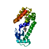
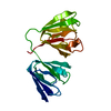
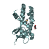
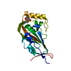
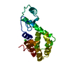
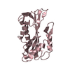
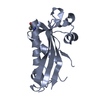
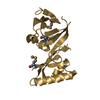
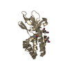
| Title | The N-terminal domain of beta-B2-crystallin resembles the putative ancestral homodimer | ||||||
 Components Components | BETA-CRYSTALLIN B2 | ||||||
 Keywords Keywords | STRUCTURAL PROTEIN / EYE LENS PROTEIN / DOMAIN INTERACTIONS / 2-FOLD SYMMETRY | ||||||
| Function / homology |  Function and homology information Function and homology informationstructural constituent of eye lens / lens development in camera-type eye / camera-type eye development / visual perception / identical protein binding Similarity search - Function | ||||||
| Biological species |  | ||||||
| Method |  X-RAY DIFFRACTION / SYNCHROTRON / MOLECULAR REPLACEMENT / Resolution: 2.35 Å X-RAY DIFFRACTION / SYNCHROTRON / MOLECULAR REPLACEMENT / Resolution: 2.35 Å | ||||||
 Authors Authors | Clout, N.J. / Basak, A. / Wieligmann, K. / Bateman, O.A. / Jaenicke, R. / Slingsby, C. | ||||||
 Citation Citation | Journal: J.Mol.Biol. / Year: 2000 Title: The N-Terminal Domain of Betab2-Crystallin Resembles the Putative Ancestral Homodimer. Authors: Clout, N.J. / Basak, A. / Wieligmann, K. / Bateman, O.A. / Jaenicke, R. / Slingsby, C. | ||||||
| History |
| ||||||
| Remark 700 | SHEET DETERMINATION METHOD: AUTHOR PROVIDED. |
- Structure visualization
| Structure viewer | Molecule: MolmilJmol/JSmol |
|---|
- Downloads & links
-Download
| PDBx/mmCIF format | 1e7n.cif.gz | 47.1 KB | Display | PDBx/mmCIF format |
|---|---|---|---|---|
| PDB format | pdb1e7n.ent.gz | 33.8 KB | Display | PDB format |
| PDBx/mmJSON format | 1e7n.json.gz | Tree view | PDBx/mmJSON format | |
| Others |  Other downloads Other downloads |
-Validation report
| Arichive directory | https://data.pdbj.org/pub/pdb/validation_reports/e7/1e7nftp://data.pdbj.org/pub/pdb/validation_reports/e7/1e7n | HTTPS FTP |
|---|
-Related structure data
| Similar structure data |
|---|

-Links
 PDBj
PDBj
- Assembly
| Deposited unit | 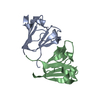
| ||||||||
|---|---|---|---|---|---|---|---|---|---|
| 1 |
| ||||||||
| Unit cell |
| ||||||||
| Noncrystallographic symmetry (NCS) | NCS oper: (Code: given Matrix: (-0.178, 0.984, -0.004), Vector: |
-Components
| #1: Protein | Mass: 11911.123 Da / Num. of mol.: 2 / Fragment: N-TERMINAL DOMAIN, RESIDUES 2-107 Source method: isolated from a genetically manipulated source Source: (gene. exp.)  #2: Water | ChemComp-HOH / |  Mass: 18.015 Da / Num. of mol.: 61 / Source method: isolated from a natural source / Formula: H2O Mass: 18.015 Da / Num. of mol.: 61 / Source method: isolated from a natural source / Formula: H2OCompound details | DOMINANT COMPONENT OF EYE LENS IN VERTEBRATE | Sequence details | ONLY N-TERMINAL DOMAIN PRESENT. | |
|---|
-Experimental details
-Experiment
| Experiment | Method: X-RAY DIFFRACTION / Number of used crystals: 1 |
|---|
- Sample preparation
| Crystal | Density Matthews: 2.23 Å3/Da / Density % sol: 45 % | |||||||||||||||||||||||||
|---|---|---|---|---|---|---|---|---|---|---|---|---|---|---|---|---|---|---|---|---|---|---|---|---|---|---|
| Crystal grow | pH: 6.5 / Details: pH 6.50 | |||||||||||||||||||||||||
| Crystal grow | *PLUS Temperature: 4 ℃ / Method: vapor diffusion, hanging dropDetails: drop consists of equal amounts of protein and reservoir solutions | |||||||||||||||||||||||||
| Components of the solutions | *PLUS
|
-Data collection
| Diffraction | Mean temperature: 100 K |
|---|---|
| Diffraction source | Source: SYNCHROTRON / Site: Photon Factory  / Type: PHOTON FACTORY / Wavelength: 0.96 / Type: PHOTON FACTORY / Wavelength: 0.96 |
| Detector | Type: MARRESEARCH / Detector: IMAGE PLATE |
| Radiation | Protocol: SINGLE WAVELENGTH / Monochromatic (M) / Laue (L): M / Scattering type: x-ray |
| Radiation wavelength | Wavelength: 0.96 Å / Relative weight: 1 |
| Reflection | Resolution: 2.35→20 Å / Num. obs: 9079 / % possible obs: 99.8 % / Redundancy: 2.7 % / Biso Wilson estimate: 16.2 Å2 / Rmerge(I) obs: 0.057 |
| Reflection | *PLUS Rmerge(I) obs: 0.05 |
- Processing
| Software |
| ||||||||||||||||||||||||||||||||||||||||||||||||||||||||||||
|---|---|---|---|---|---|---|---|---|---|---|---|---|---|---|---|---|---|---|---|---|---|---|---|---|---|---|---|---|---|---|---|---|---|---|---|---|---|---|---|---|---|---|---|---|---|---|---|---|---|---|---|---|---|---|---|---|---|---|---|---|---|
| Refinement | Method to determine structure: MOLECULAR REPLACEMENT / Resolution: 2.35→19.89 Å / Rfactor Rfree error: 0.012 / Cross valid method: THROUGHOUT / σ(F): 0 Details: THE N-TERMINAL 13 RESIDUE EXTENSION WAS NOT SEEN IN THE DENSITY C-TERMINAL LAST FOUR RESIDUES WERE NOT SEEN IN THE DENSITY MAPS
| ||||||||||||||||||||||||||||||||||||||||||||||||||||||||||||
| Displacement parameters | Biso mean: 21.6 Å2
| ||||||||||||||||||||||||||||||||||||||||||||||||||||||||||||
| Refine analyze |
| ||||||||||||||||||||||||||||||||||||||||||||||||||||||||||||
| Refinement step | Cycle: LAST / Resolution: 2.35→19.89 Å
| ||||||||||||||||||||||||||||||||||||||||||||||||||||||||||||
| Refine LS restraints |
| ||||||||||||||||||||||||||||||||||||||||||||||||||||||||||||
| LS refinement shell | Resolution: 2.35→2.5 Å / Rfactor Rfree error: 0.034 / Total num. of bins used: 6
| ||||||||||||||||||||||||||||||||||||||||||||||||||||||||||||
| Software | *PLUS Name: CNS / Version: 0.9 / Classification: refinement | ||||||||||||||||||||||||||||||||||||||||||||||||||||||||||||
| Refinement | *PLUS Rfactor Rwork: 0.2049 | ||||||||||||||||||||||||||||||||||||||||||||||||||||||||||||
| Solvent computation | *PLUS | ||||||||||||||||||||||||||||||||||||||||||||||||||||||||||||
| Displacement parameters | *PLUS | ||||||||||||||||||||||||||||||||||||||||||||||||||||||||||||
| Refine LS restraints | *PLUS
|
