 Movie
Movie Controller


+ Open data

- Basic information
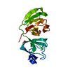
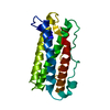
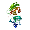
| Entry | Database: PDB / ID: 1hk0 | ||||||
|---|---|---|---|---|---|---|---|
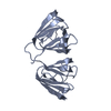
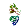
| Title | Human GammaD Crystallin Structure at 1.25 A Resolution | ||||||
 Components Components | Gamma-crystallin D | ||||||
 Keywords Keywords | EYE LENS PROTEIN / CATARACT | ||||||
| Function / homology |  Function and homology information Function and homology informationlens fiber cell differentiation / structural constituent of eye lens / lens development in camera-type eye / visual perception / cellular response to reactive oxygen species / nucleus / cytoplasm Similarity search - Function | ||||||
| Biological species |  Homo sapiens (human) Homo sapiens (human) | ||||||
| Method |  X-RAY DIFFRACTION / SYNCHROTRON / MOLECULAR REPLACEMENT / Resolution: 1.25 Å X-RAY DIFFRACTION / SYNCHROTRON / MOLECULAR REPLACEMENT / Resolution: 1.25 Å | ||||||
 Authors Authors | Basak, A.K. / Slingsby, C. | ||||||
 Citation Citation | Journal: J.Mol.Biol. / Year: 2003 Title: High-Resolution X-Ray Crystal Structures of Human Gammad Crystallin (1.25A) and the R58H Mutant (1.15A) Associated with Aculeiform Cataract Authors: Basak, A.K. / Bateman, O. / Slingsby, C. / Pande, A. / Asherie, N. / Ogun, O. / Benedek, G. / Pande, J. #1: Journal: Exp.Eye Res. / Year: 1991 Title: Crystal Structure of Calf Eye Lens Gamma-Crystallin Iiib at 2.5 A Resolution: Its Relation to Function Authors: Yu, C. / Nevskaya, N. / Vernoslova, E. / Nikonov, S. / Yu, S. / Brazhnikov, E. / Fomenkova, N. / Lunin, V. / Urzhumtsev, A. | ||||||
| History |
| ||||||
| Remark 700 | SHEET THE SHEET STRUCTURE OF THIS MOLECULE IS BIFURCATED. IN ORDER TO REPRESENT THIS FEATURE IN ... SHEET THE SHEET STRUCTURE OF THIS MOLECULE IS BIFURCATED. IN ORDER TO REPRESENT THIS FEATURE IN THE SHEET RECORDS BELOW, TWO SHEETS ARE DEFINED. |
- Structure visualization
| Structure viewer | Molecule: MolmilJmol/JSmol |
|---|
- Downloads & links
-Download
| PDBx/mmCIF format | 1hk0.cif.gz | 95.9 KB | Display | PDBx/mmCIF format |
|---|---|---|---|---|
| PDB format | pdb1hk0.ent.gz | 72.3 KB | Display | PDB format |
| PDBx/mmJSON format | 1hk0.json.gz | Tree view | PDBx/mmJSON format | |
| Others |  Other downloads Other downloads |
-Validation report
| Arichive directory | https://data.pdbj.org/pub/pdb/validation_reports/hk/1hk0ftp://data.pdbj.org/pub/pdb/validation_reports/hk/1hk0 | HTTPS FTP |
|---|
-Related structure data
| Related structure data | 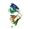 1h4aC 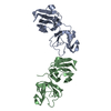 1elpS C: citing same article ( S: Starting model for refinement |
|---|---|
| Similar structure data |


-Links
 PDBj
PDBj
- Assembly
| Deposited unit | 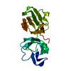
| ||||||||
|---|---|---|---|---|---|---|---|---|---|
| 1 |
| ||||||||
| Unit cell |
|
-Components
| #1: Protein | Mass: 20634.969 Da / Num. of mol.: 1 Source method: isolated from a genetically manipulated source Source: (gene. exp.) Homo sapiens (human) / Gene: CRYGD, CRYG4 / Production host:  |
|---|---|
| #2: Water | ChemComp-HOH /  Mass: 18.015 Da / Num. of mol.: 282 / Source method: isolated from a natural source / Formula: H2O Mass: 18.015 Da / Num. of mol.: 282 / Source method: isolated from a natural source / Formula: H2O |
-Experimental details
-Experiment
| Experiment | Method: X-RAY DIFFRACTION / Number of used crystals: 1 |
|---|
- Sample preparation
| Crystal | Density Matthews: 1.87 Å3/Da / Density % sol: 38 % | ||||||||||||||||||
|---|---|---|---|---|---|---|---|---|---|---|---|---|---|---|---|---|---|---|---|
| Crystal grow | Temperature: 277 K / pH: 7 / Details: 10MM PHOSPHATE, 4 DEGREE CELSIUS, pH 7.00 | ||||||||||||||||||
| Crystal grow | *PLUS pH: 7 / Method: batch method | ||||||||||||||||||
| Components of the solutions | *PLUS
|
-Data collection
| Diffraction | Mean temperature: 100 K |
|---|---|
| Diffraction source | Source: SYNCHROTRON / Site: ESRF  / Beamline: ID14-1 / Wavelength: 0.933 / Beamline: ID14-1 / Wavelength: 0.933 |
| Detector | Type: MARRESEARCH / Detector: CCD / Date: Apr 15, 2001 / Details: MIRROR |
| Radiation | Monochromator: LAUE DIAMOND C111 / Protocol: SINGLE WAVELENGTH / Monochromatic (M) / Laue (L): M / Scattering type: x-ray |
| Radiation wavelength | Wavelength: 0.933 Å / Relative weight: 1 |
| Reflection | Resolution: 1.25→28.17 Å / Num. obs: 39066 / % possible obs: 86.8 % / Redundancy: 2.9 % / Rmerge(I) obs: 0.048 / Net I/σ(I): 7.4 |
| Reflection shell | Resolution: 1.25→1.32 Å / Redundancy: 1.6 % / Rmerge(I) obs: 0.189 / Mean I/σ(I) obs: 3.7 / % possible all: 45.5 |
| Reflection | *PLUS Num. obs: 39021 / Num. measured all: 212732 |
| Reflection shell | *PLUS % possible obs: 45.5 % / Redundancy: 1.9 % |
- Processing
| Software |
| ||||||||||||||||||||
|---|---|---|---|---|---|---|---|---|---|---|---|---|---|---|---|---|---|---|---|---|---|
| Refinement | Method to determine structure: MOLECULAR REPLACEMENT Starting model: PDB ENTRY 1ELP Resolution: 1.25→28.17 Å / SU B: 0.877 / SU ML: 0.038 / Cross valid method: THROUGHOUT / σ(F): 0 / ESU R: 0.066 / ESU R Free: 0.062 / Details: HYDROGENS HAVE BEEN ADDED IN THE RIDING POSITIONS
| ||||||||||||||||||||
| Displacement parameters | Biso mean: 13.5 Å2
| ||||||||||||||||||||
| Refinement step | Cycle: LAST / Resolution: 1.25→28.17 Å
| ||||||||||||||||||||
| Refinement | *PLUS % reflection Rfree: 8 % / Rfactor obs: 0.1671 / Rfactor Rfree: 0.2078 / Rfactor Rwork: 0.1636 | ||||||||||||||||||||
| Solvent computation | *PLUS | ||||||||||||||||||||
| Displacement parameters | *PLUS | ||||||||||||||||||||
| Refine LS restraints | *PLUS
|
