 Movie
Movie Controller


+ Open data

- Basic information
| Entry | Database: PDB / ID: 1zie | ||||||
|---|---|---|---|---|---|---|---|
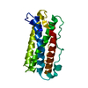
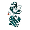
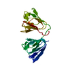
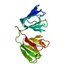
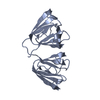
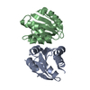
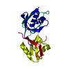
| Title | Hydrogenated gammaE crystallin in D2O solvent | ||||||
 Components Components | Gamma crystallin E | ||||||
 Keywords Keywords | STRUCTURAL PROTEIN / 4 Greek key motifs | ||||||
| Function / homology |  Function and homology information Function and homology informationstructural constituent of eye lens / lens development in camera-type eye / visual perception Similarity search - Function | ||||||
| Biological species |  | ||||||
| Method |  X-RAY DIFFRACTION / SYNCHROTRON / MOLECULAR REPLACEMENT / Resolution: 1.44 Å X-RAY DIFFRACTION / SYNCHROTRON / MOLECULAR REPLACEMENT / Resolution: 1.44 Å | ||||||
 Authors Authors | Artero, J.B. / Hartlein, M. / McSweeney, S. / Timmins, P. | ||||||
 Citation Citation | Journal: Acta Crystallogr.,Sect.D / Year: 2005 Title: A comparison of refined X-ray structures of hydrogenated and perdeuterated rat gammaE-crystallin in H2O and D2O. Authors: Artero, J.B. / Hartlein, M. / McSweeney, S. / Timmins, P. | ||||||
| History |
|
- Structure visualization
| Structure viewer | Molecule: MolmilJmol/JSmol |
|---|
- Downloads & links
-Download
| PDBx/mmCIF format | 1zie.cif.gz | 57.5 KB | Display | PDBx/mmCIF format |
|---|---|---|---|---|
| PDB format | pdb1zie.ent.gz | 40.9 KB | Display | PDB format |
| PDBx/mmJSON format | 1zie.json.gz | Tree view | PDBx/mmJSON format | |
| Others |  Other downloads Other downloads |
-Validation report
| Arichive directory | https://data.pdbj.org/pub/pdb/validation_reports/zi/1zieftp://data.pdbj.org/pub/pdb/validation_reports/zi/1zie | HTTPS FTP |
|---|
-Related structure data
| Related structure data |  1zgtC  1ziqC  1zirC  1a5dS S: Starting model for refinement C: citing same article ( |
|---|---|
| Similar structure data |


-Links
 PDBj
PDBj


- Assembly
| Deposited unit | 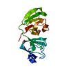
| ||||||||
|---|---|---|---|---|---|---|---|---|---|
| 1 |
| ||||||||
| Unit cell |
|
-Components
| #1: Protein | Mass: 21163.611 Da / Num. of mol.: 1 Source method: isolated from a genetically manipulated source Source: (gene. exp.)  | ||
|---|---|---|---|
| #2: Chemical |   Mass: 59.044 Da / Num. of mol.: 2 / Source method: obtained synthetically / Formula: C2H3O2 Mass: 59.044 Da / Num. of mol.: 2 / Source method: obtained synthetically / Formula: C2H3O2#3: Chemical | ChemComp-DOD / |   Mass: 18.015 Da / Num. of mol.: 202 / Source method: isolated from a natural source / Formula: D2O Mass: 18.015 Da / Num. of mol.: 202 / Source method: isolated from a natural source / Formula: D2O |
-Experimental details
-Experiment
| Experiment | Method: X-RAY DIFFRACTION / Number of used crystals: 1 |
|---|
- Sample preparation
| Crystal | Density Matthews: 2 Å3/Da / Density % sol: 37.44 % |
|---|---|
| Crystal grow | Temperature: 293 K / Method: vapor diffusion, hanging drop / pH: 7.4 Details: PEG 3350, Magnesium Acetate, D2O, pH 7.4, VAPOR DIFFUSION, HANGING DROP, temperature 293K |
-Data collection
| Diffraction | Mean temperature: 100 K |
|---|---|
| Diffraction source | Source: SYNCHROTRON / Site: ESRF  / Beamline: ID14-4 / Wavelength: 0.9393 Å / Beamline: ID14-4 / Wavelength: 0.9393 Å |
| Detector | Type: ADSC QUANTUM 4 / Detector: CCD / Date: Mar 11, 2004 |
| Radiation | Monochromator: a double crystal monochromator / Protocol: SINGLE WAVELENGTH / Monochromatic (M) / Laue (L): M / Scattering type: x-ray |
| Radiation wavelength | Wavelength: 0.9393 Å / Relative weight: 1 |
| Reflection | Resolution: 1.44→50.64 Å / Num. all: 30894 / Num. obs: 30005 / % possible obs: 97.1 % / Observed criterion σ(F): 2 / Observed criterion σ(I): 2 |
| Reflection shell | Resolution: 1.44→1.53 Å / % possible all: 90.3 |
- Processing
| Software |
| ||||||||||||||||||||
|---|---|---|---|---|---|---|---|---|---|---|---|---|---|---|---|---|---|---|---|---|---|
| Refinement | Method to determine structure: MOLECULAR REPLACEMENT Starting model: 1A5D Resolution: 1.44→50.64 Å / σ(F): 4 / Stereochemistry target values: Engh & Huber
| ||||||||||||||||||||
| Refinement step | Cycle: LAST / Resolution: 1.44→50.64 Å
| ||||||||||||||||||||
| Refine LS restraints |
|
