 Movie
Movie Controller

[English] 日本語
 Yorodumi
- PDB-1dcn: INACTIVE MUTANT H162N OF DELTA 2 CRYSTALLIN WITH BOUND ARGININOSU... -
+ Open data

- Basic information
| Entry | Database: PDB / ID: 1dcn | ||||||
|---|---|---|---|---|---|---|---|
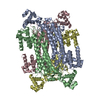
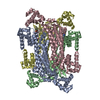
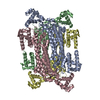
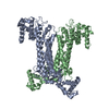
| Title | INACTIVE MUTANT H162N OF DELTA 2 CRYSTALLIN WITH BOUND ARGININOSUCCINATE | ||||||
 Components Components | DELTA 2 CRYSTALLIN | ||||||
 Keywords Keywords | EYE LENS PROTEIN / DELTA 2 CRYSTALLIN / ARGININOSUCCINATE LYASE | ||||||
| Function / homology |  Function and homology information Function and homology informationargininosuccinate lyase / argininosuccinate lyase activity / : / structural constituent of eye lens / L-arginine biosynthetic process / cytosol Similarity search - Function | ||||||
| Biological species |  | ||||||
| Method |  X-RAY DIFFRACTION / SYNCHROTRON / MOLECULAR REPLACEMENT / Resolution: 2.3 Å X-RAY DIFFRACTION / SYNCHROTRON / MOLECULAR REPLACEMENT / Resolution: 2.3 Å | ||||||
 Authors Authors | Vallee, F. / Turner, M.A. / Lindley, P. / Howell, P.L. | ||||||
 Citation Citation | Journal: Biochemistry / Year: 1999 Title: Crystal structure of an inactive duck delta II crystallin mutant with bound argininosuccinate. Authors: Vallee, F. / Turner, M.A. / Lindley, P.L. / Howell, P.L. | ||||||
| History |
|
- Structure visualization
| Structure viewer | Molecule: MolmilJmol/JSmol |
|---|
- Downloads & links
-Download
| PDBx/mmCIF format | 1dcn.cif.gz | 338.8 KB | Display | PDBx/mmCIF format |
|---|---|---|---|---|
| PDB format | pdb1dcn.ent.gz | 278.1 KB | Display | PDB format |
| PDBx/mmJSON format | 1dcn.json.gz | Tree view | PDBx/mmJSON format | |
| Others |  Other downloads Other downloads |
-Validation report
| Arichive directory | https://data.pdbj.org/pub/pdb/validation_reports/dc/1dcnftp://data.pdbj.org/pub/pdb/validation_reports/dc/1dcn | HTTPS FTP |
|---|
-Related structure data
| Related structure data |  1auwS S: Starting model for refinement |
|---|---|
| Similar structure data |


-Links
 PDBj
PDBj

- Assembly
| Deposited unit | 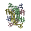
| ||||||||||||||||
|---|---|---|---|---|---|---|---|---|---|---|---|---|---|---|---|---|---|
| 1 |
| ||||||||||||||||
| Unit cell |
| ||||||||||||||||
| Noncrystallographic symmetry (NCS) | NCS oper:
|
-Components
| #1: Protein | Mass: 49500.797 Da / Num. of mol.: 4 / Mutation: H162N Source method: isolated from a genetically manipulated source Details: ARGININOSUCCINATE BOUND TO ONE ACTIVE SITE / Source: (gene. exp.)  #2: Chemical |   Mass: 290.273 Da / Num. of mol.: 1 / Source method: obtained synthetically / Formula: C10H18N4O6 / Details: ARGININOSUCCINATE BOUND TO ONE ACTIVE SITE Mass: 290.273 Da / Num. of mol.: 1 / Source method: obtained synthetically / Formula: C10H18N4O6 / Details: ARGININOSUCCINATE BOUND TO ONE ACTIVE SITE#3: Water | ChemComp-HOH / |  Mass: 18.015 Da / Num. of mol.: 471 / Source method: isolated from a natural source / Formula: H2O / Details: ARGININOSUCCINATE BOUND TO ONE ACTIVE SITE Mass: 18.015 Da / Num. of mol.: 471 / Source method: isolated from a natural source / Formula: H2O / Details: ARGININOSUCCINATE BOUND TO ONE ACTIVE SITE |
|---|
-Experimental details
-Experiment
| Experiment | Method: X-RAY DIFFRACTION |
|---|
- Sample preparation
| Crystal | Density Matthews: 2.5 Å3/Da / Density % sol: 50.7 % | ||||||||||||||||||||||||||||||||||||
|---|---|---|---|---|---|---|---|---|---|---|---|---|---|---|---|---|---|---|---|---|---|---|---|---|---|---|---|---|---|---|---|---|---|---|---|---|---|
| Crystal grow | pH: 8.5 / Details: pH 8.5 | ||||||||||||||||||||||||||||||||||||
| Crystal grow | *PLUS Method: vapor diffusion, hanging drop | ||||||||||||||||||||||||||||||||||||
| Components of the solutions | *PLUS
|
-Data collection
| Diffraction | Mean temperature: 120 K |
|---|---|
| Diffraction source | Source: SYNCHROTRON / Site: CHESS  / Beamline: F1 / Wavelength: 0.91 / Beamline: F1 / Wavelength: 0.91 |
| Radiation | Monochromatic (M) / Laue (L): M / Scattering type: x-ray |
| Radiation wavelength | Wavelength: 0.91 Å / Relative weight: 1 |
| Reflection | Resolution: 2.3→20 Å / Num. obs: 77291 / % possible obs: 90.3 % / Observed criterion σ(I): 0 / Redundancy: 2.7 % / Biso Wilson estimate: 27.5 Å2 / Rsym value: 0.079 |
| Reflection shell | Resolution: 2.3→2.38 Å / Rmerge(I) obs: 0.233 |
| Reflection | *PLUS Num. measured all: 194258 / Rmerge(I) obs: 0.079 |
| Reflection shell | *PLUS Highest resolution: 2.3 Å / % possible obs: 96.4 % |
- Processing
| Software |
| ||||||||||||||||||||||||||||||||||||||||||||||||||||||||||||
|---|---|---|---|---|---|---|---|---|---|---|---|---|---|---|---|---|---|---|---|---|---|---|---|---|---|---|---|---|---|---|---|---|---|---|---|---|---|---|---|---|---|---|---|---|---|---|---|---|---|---|---|---|---|---|---|---|---|---|---|---|---|
| Refinement | Method to determine structure: MOLECULAR REPLACEMENT Starting model: 1AUW Resolution: 2.3→20 Å / Rfactor Rfree error: 0.003 / Data cutoff high rms absF: 9791538.7 / Isotropic thermal model: RESTRAINED / Cross valid method: THROUGHOUT / σ(F): 0
| ||||||||||||||||||||||||||||||||||||||||||||||||||||||||||||
| Solvent computation | Solvent model: FLAT MODEL / Bsol: 61.84 Å2 / ksol: 0.346 e/Å3 | ||||||||||||||||||||||||||||||||||||||||||||||||||||||||||||
| Displacement parameters | Biso mean: 46.3 Å2
| ||||||||||||||||||||||||||||||||||||||||||||||||||||||||||||
| Refine analyze |
| ||||||||||||||||||||||||||||||||||||||||||||||||||||||||||||
| Refinement step | Cycle: LAST / Resolution: 2.3→20 Å
| ||||||||||||||||||||||||||||||||||||||||||||||||||||||||||||
| Refine LS restraints |
| ||||||||||||||||||||||||||||||||||||||||||||||||||||||||||||
| LS refinement shell | Resolution: 2.3→2.44 Å / Rfactor Rfree error: 0.011 / Total num. of bins used: 6
| ||||||||||||||||||||||||||||||||||||||||||||||||||||||||||||
| Xplor file |
| ||||||||||||||||||||||||||||||||||||||||||||||||||||||||||||
| Software | *PLUS Name: CNS / Version: 0.3C / Classification: refinement | ||||||||||||||||||||||||||||||||||||||||||||||||||||||||||||
| Refinement | *PLUS Lowest resolution: 17 Å / Rfactor Rfree: 0.29 | ||||||||||||||||||||||||||||||||||||||||||||||||||||||||||||
| Solvent computation | *PLUS | ||||||||||||||||||||||||||||||||||||||||||||||||||||||||||||
| Displacement parameters | *PLUS | ||||||||||||||||||||||||||||||||||||||||||||||||||||||||||||
| Refine LS restraints | *PLUS
|
