 Movie
Movie Controller


+ Open data

- Basic information
| Entry | Database: PDB / ID: 1k7w | ||||||
|---|---|---|---|---|---|---|---|
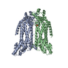
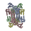
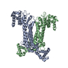
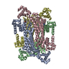
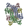
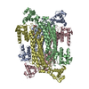
| Title | Crystal Structure of S283A Duck Delta 2 Crystallin Mutant | ||||||
 Components Components | delta 2 crystallin | ||||||
 Keywords Keywords | LYASE / eye lens protein / delta 2 crystallin / argininosuccinate lyase / enzyme mechanism | ||||||
| Function / homology |  Function and homology information Function and homology informationargininosuccinate lyase / argininosuccinate lyase activity / : / structural constituent of eye lens / L-arginine biosynthetic process / cytosol Similarity search - Function | ||||||
| Biological species |  | ||||||
| Method |  X-RAY DIFFRACTION / SYNCHROTRON / MOLECULAR REPLACEMENT / Resolution: 1.96 Å X-RAY DIFFRACTION / SYNCHROTRON / MOLECULAR REPLACEMENT / Resolution: 1.96 Å | ||||||
 Authors Authors | Sampaleanu, L.M. / Yu, B. / Howell, P.L. | ||||||
 Citation Citation | Journal: J.Biol.Chem. / Year: 2002 Title: Mutational analysis of duck delta 2 crystallin and the structure of an inactive mutant with bound substrate provide insight into the enzymatic mechanism of argininosuccinate lyase. Authors: Sampaleanu, L.M. / Yu, B. / Howell, P.L. | ||||||
| History |
|
- Structure visualization
| Structure viewer | Molecule: MolmilJmol/JSmol |
|---|
- Downloads & links
-Download
| PDBx/mmCIF format | 1k7w.cif.gz | 358.8 KB | Display | PDBx/mmCIF format |
|---|---|---|---|---|
| PDB format | pdb1k7w.ent.gz | 293.8 KB | Display | PDB format |
| PDBx/mmJSON format | 1k7w.json.gz | Tree view | PDBx/mmJSON format | |
| Others |  Other downloads Other downloads |
-Validation report
| Arichive directory | https://data.pdbj.org/pub/pdb/validation_reports/k7/1k7wftp://data.pdbj.org/pub/pdb/validation_reports/k7/1k7w | HTTPS FTP |
|---|
-Related structure data
| Related structure data | 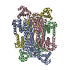 1hy1S S: Starting model for refinement |
|---|---|
| Similar structure data |


-Links
 PDBj
PDBj

- Assembly
| Deposited unit | 
| ||||||||
|---|---|---|---|---|---|---|---|---|---|
| 1 |
| ||||||||
| Unit cell |
| ||||||||
| Details | The biological assembly is the homotetramer with four bound argininosuccinate molecules, as present in the asymmetric unit |
-Components
| #1: Protein | Mass: 51743.355 Da / Num. of mol.: 4 / Mutation: S283A Source method: isolated from a genetically manipulated source Source: (gene. exp.)  #2: Chemical | ChemComp-AS1 /   Mass: 290.273 Da / Num. of mol.: 4 / Source method: obtained synthetically / Formula: C10H18N4O6 Mass: 290.273 Da / Num. of mol.: 4 / Source method: obtained synthetically / Formula: C10H18N4O6#3: Water | ChemComp-HOH / |  Mass: 18.015 Da / Num. of mol.: 707 / Source method: isolated from a natural source / Formula: H2O Mass: 18.015 Da / Num. of mol.: 707 / Source method: isolated from a natural source / Formula: H2O |
|---|
-Experimental details
-Experiment
| Experiment | Method: X-RAY DIFFRACTION / Number of used crystals: 1 |
|---|
- Sample preparation
| Crystal | Density Matthews: 2.32 Å3/Da / Density % sol: 47.1 % | ||||||||||||||||||||||||||||||||||||||||||||||||||||||||
|---|---|---|---|---|---|---|---|---|---|---|---|---|---|---|---|---|---|---|---|---|---|---|---|---|---|---|---|---|---|---|---|---|---|---|---|---|---|---|---|---|---|---|---|---|---|---|---|---|---|---|---|---|---|---|---|---|---|
| Crystal grow | Temperature: 298 K / Method: vapor diffusion, hanging drop / pH: 7.5 Details: 12% PEG 2000 MME, 300 mM magnesium chloride, 100 mM HEPES, pH 7.5, VAPOR DIFFUSION, HANGING DROP, temperature 298K | ||||||||||||||||||||||||||||||||||||||||||||||||||||||||
| Crystal grow | *PLUS pH: 7.4 | ||||||||||||||||||||||||||||||||||||||||||||||||||||||||
| Components of the solutions | *PLUS
|
-Data collection
| Diffraction | Mean temperature: 100 K |
|---|---|
| Diffraction source | Source: SYNCHROTRON / Site: NSLS  / Beamline: X8C / Wavelength: 0.96 Å / Beamline: X8C / Wavelength: 0.96 Å |
| Detector | Type: MARRESEARCH / Detector: IMAGE PLATE / Date: May 18, 2000 |
| Radiation | Monochromator: parabolic collimating mirror placed upstream of crystal monochromator Protocol: SINGLE WAVELENGTH / Monochromatic (M) / Laue (L): M / Scattering type: x-ray |
| Radiation wavelength | Wavelength: 0.96 Å / Relative weight: 1 |
| Reflection | Resolution: 1.96→20 Å / Num. all: 125073 / Num. obs: 125073 / % possible obs: 94 % / Observed criterion σ(F): 0 / Observed criterion σ(I): 0 / Redundancy: 5.4 % / Biso Wilson estimate: 16.8 Å2 / Rsym value: 0.08 / Net I/σ(I): 10.9 |
| Reflection shell | Resolution: 1.96→2.03 Å / Rsym value: 0.34 / % possible all: 91.8 |
| Reflection | *PLUS Highest resolution: 1.94 Å / Lowest resolution: 20 Å / Num. obs: 127416 / % possible obs: 94 % / Num. measured all: 692205 / Rmerge(I) obs: 0.08 |
| Reflection shell | *PLUS % possible obs: 91.8 % / Rmerge(I) obs: 0.34 |
- Processing
| Software |
| |||||||||||||||||||||||||
|---|---|---|---|---|---|---|---|---|---|---|---|---|---|---|---|---|---|---|---|---|---|---|---|---|---|---|
| Refinement | Method to determine structure: MOLECULAR REPLACEMENT Starting model: 1HY1 Resolution: 1.96→19.81 Å / Rfactor Rfree error: 0.002 / Data cutoff high absF: 299780.83 / Data cutoff low absF: 0 / Isotropic thermal model: RESTRAINED / Cross valid method: THROUGHOUT / σ(F): 0 / σ(I): 0 / Stereochemistry target values: Engh & Huber
| |||||||||||||||||||||||||
| Solvent computation | Solvent model: FLAT MODEL / Bsol: 57.7155 Å2 / ksol: 0.393911 e/Å3 | |||||||||||||||||||||||||
| Displacement parameters | Biso mean: 30 Å2
| |||||||||||||||||||||||||
| Refine analyze |
| |||||||||||||||||||||||||
| Refinement step | Cycle: LAST / Resolution: 1.96→19.81 Å
| |||||||||||||||||||||||||
| Refine LS restraints |
| |||||||||||||||||||||||||
| LS refinement shell | Resolution: 1.96→2.08 Å / Rfactor Rfree error: 0.007 / Total num. of bins used: 6
| |||||||||||||||||||||||||
| Xplor file |
| |||||||||||||||||||||||||
| Refinement | *PLUS Lowest resolution: 20 Å / σ(F): 0 / % reflection Rfree: 10 % / Rfactor obs: 0.207 | |||||||||||||||||||||||||
| Solvent computation | *PLUS | |||||||||||||||||||||||||
| Displacement parameters | *PLUS Biso mean: 30 Å2 | |||||||||||||||||||||||||
| Refine LS restraints | *PLUS
| |||||||||||||||||||||||||
| LS refinement shell | *PLUS Lowest resolution: 2.03 Å / Rfactor Rfree: 0.301 / % reflection Rfree: 9.9 % / Rfactor Rwork: 0.257 |
