 Movie
Movie Controller


+ Open data

- Basic information
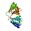
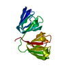
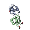
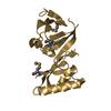
| Entry | Database: PDB / ID: 1gcs | ||||||
|---|---|---|---|---|---|---|---|
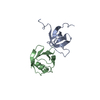
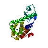
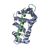
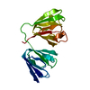
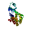
| Title | STRUCTURE OF THE BOVINE GAMMA-B CRYSTALLIN AT 150K | ||||||
 Components Components | GAMMA-B CRYSTALLIN | ||||||
 Keywords Keywords | EYE LENS PROTEIN | ||||||
| Function / homology |  Function and homology information Function and homology informationstructural constituent of eye lens / lens development in camera-type eye / visual perception Similarity search - Function | ||||||
| Biological species |  | ||||||
| Method |  X-RAY DIFFRACTION / Resolution: 2 Å X-RAY DIFFRACTION / Resolution: 2 Å | ||||||
 Authors Authors | Najmudin, S. / Lindley, P. / Slingsby, C. / Bateman, O. / Myles, D. / Kumaraswamy, S. / Glover, I. | ||||||
 Citation Citation | Journal: J.CHEM.SOC.,FARADAY TRANS. / Year: 1993 Title: Structure of the Bovine Gamma-B Crystallin at 150K Authors: Lindley, P.F. / Najmudin, S. / Bateman, O. / Slingsby, C. / Myles, D. / Kumaraswamy, S. / Glover, I. #1: Journal: Acta Crystallogr.,Sect.D / Year: 1993Title: Lindley Structure of Bovine Gamma-B (Gamma-II) Crystallin at 1.47 Angstroms Resolution Authors: Najmudin, S. / Nalini, V. / Driessen, H.P.C. / Slingsby, C. / Blundell, T.L. / Moss, D.S. / Lindley, P.F. #2: Journal: Pept.Protein Rev. / Year: 1984Title: X-Ray Studies of the Lens Specific Proteins, the Crystallins Authors: Summers, L. / Wistow, G. / Narebor, M. / Moss, D. / Lindley, P. / Slingsby, C. / Blundell, T. / Bartunik, H. / Bartels, K. #3: Journal: J.Mol.Biol. / Year: 1983Title: X-Ray Analysis of the Eye Lens Protein Gamma-II Crystallin at 1.9 Angstroms Resolution Authors: Wistow, G. / Turnell, B. / Summers, L. / Slingsby, C. / Moss, D. / Miller, L. / Lindley, P. / Blundell, T. #4: Journal: Nature / Year: 1981Title: The Molecular Structure and Stability of the Eye Lens: X-Ray Analysis of Gamma Crystallin II Authors: Blundell, T. / Lindley, P. / Miller, L. / Moss, D. / Slingsby, C. / Tickle, I. / Turnell, B. / Wistow, G. | ||||||
| History |
| ||||||
| Remark 700 | SHEET IN *SHEET* RECORDS BELOW, THERE IS A POSSIBLE BETA-BRIDGE BETWEEN RESIDUES 119 - 121 AND 162 - 164. |
- Structure visualization
| Structure viewer | Molecule: MolmilJmol/JSmol |
|---|
- Downloads & links
-Download
| PDBx/mmCIF format | 1gcs.cif.gz | 54 KB | Display | PDBx/mmCIF format |
|---|---|---|---|---|
| PDB format | pdb1gcs.ent.gz | 39.2 KB | Display | PDB format |
| PDBx/mmJSON format | 1gcs.json.gz | Tree view | PDBx/mmJSON format | |
| Others |  Other downloads Other downloads |
-Validation report
| Arichive directory | https://data.pdbj.org/pub/pdb/validation_reports/gc/1gcsftp://data.pdbj.org/pub/pdb/validation_reports/gc/1gcs | HTTPS FTP |
|---|
-Related structure data
| Similar structure data |
|---|

-Links
 PDBj
PDBj
- Assembly
| Deposited unit | 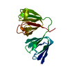
| ||||||||
|---|---|---|---|---|---|---|---|---|---|
| 1 |
| ||||||||
| Unit cell |
| ||||||||
| Atom site foot note | 1: THE SIDE CHAINS OF CYS 18 AND CYS 22 ARE IN THE FULLY REDUCED STATE. |
-Components
| #1: Protein | Mass: 20992.559 Da / Num. of mol.: 1 Source method: isolated from a genetically manipulated source Source: (gene. exp.) |
|---|---|
| #2: Water | ChemComp-HOH /  Mass: 18.015 Da / Num. of mol.: 255 / Source method: isolated from a natural source / Formula: H2O Mass: 18.015 Da / Num. of mol.: 255 / Source method: isolated from a natural source / Formula: H2O |
-Experimental details
-Experiment
| Experiment | Method: X-RAY DIFFRACTION |
|---|
- Sample preparation
| Crystal | Density Matthews: 1.84 Å3/Da / Density % sol: 33.13 % | ||||||||||||||||||||
|---|---|---|---|---|---|---|---|---|---|---|---|---|---|---|---|---|---|---|---|---|---|
| Crystal grow | *PLUS Temperature: 0 K / pH: 7 / Method: unknown / Details: Carlisle, C.H.,(1977) J. Mol. Biol., 110, 417. | ||||||||||||||||||||
| Components of the solutions | *PLUS
|
-Data collection
| Radiation | Scattering type: x-ray |
|---|---|
| Radiation wavelength | Relative weight: 1 |
| Reflection | *PLUS Highest resolution: 1.95 Å / Num. all: 11407 / Num. obs: 10735 / Num. measured all: 36656 / Rmerge(I) obs: 0.052 |
- Processing
| Software | Name: RESTRAIN / Classification: refinement | ||||||||||||
|---|---|---|---|---|---|---|---|---|---|---|---|---|---|
| Refinement | Resolution: 2→8 Å / σ(F): 0 /
| ||||||||||||
| Refinement step | Cycle: LAST / Resolution: 2→8 Å
| ||||||||||||
| Refine LS restraints |
| ||||||||||||
| Refinement | *PLUS Rfactor obs: 0.17 | ||||||||||||
| Solvent computation | *PLUS | ||||||||||||
| Displacement parameters | *PLUS |
