 Movie
Movie Controller


+ Open data

- Basic information
| Entry | Database: PDB / ID: 5wsv | ||||||
|---|---|---|---|---|---|---|---|
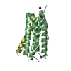
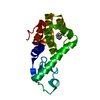
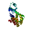
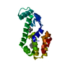
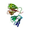
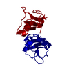
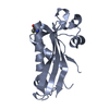
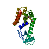
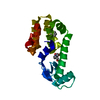
| Title | Crystal structure of Myosin VIIa IQ5 in complex with Ca2+-CaM | ||||||
 Components Components |
| ||||||
 Keywords Keywords | MOTOR PROTEIN/CALCIUM BINDING PROTEIN / Molecular motor / Calcium signaling / Protein complex / Calmodulin / MOTOR PROTEIN-CALCIUM BINDING PROTEIN complex | ||||||
| Function / homology |  Function and homology information Function and homology informationpigment granule localization / pigment granule transport / upper tip-link density / The canonical retinoid cycle in rods (twilight vision) / myosin VII complex / stereocilium base / inner ear receptor cell differentiation / phagolysosome assembly / equilibrioception / mechanoreceptor differentiation ...pigment granule localization / pigment granule transport / upper tip-link density / The canonical retinoid cycle in rods (twilight vision) / myosin VII complex / stereocilium base / inner ear receptor cell differentiation / phagolysosome assembly / equilibrioception / mechanoreceptor differentiation / sensory perception of light stimulus / inner ear receptor cell stereocilium organization / photoreceptor connecting cilium / : / : / : / : / positive regulation of protein autophosphorylation / inner ear auditory receptor cell differentiation / : / sensory organ development / negative regulation of peptidyl-threonine phosphorylation / actin filament-based movement / : / stereocilium / type 3 metabotropic glutamate receptor binding / auditory receptor cell stereocilium organization / positive regulation of peptidyl-threonine phosphorylation / myosin complex / cell projection organization / positive regulation of DNA binding / CaM pathway / Cam-PDE 1 activation / Sodium/Calcium exchangers / inner ear morphogenesis / Calmodulin induced events / Reduction of cytosolic Ca++ levels / Activation of Ca-permeable Kainate Receptor / CREB1 phosphorylation through the activation of CaMKII/CaMKK/CaMKIV cascasde / Loss of phosphorylation of MECP2 at T308 / CREB1 phosphorylation through the activation of Adenylate Cyclase / sensory perception / negative regulation of high voltage-gated calcium channel activity / PKA activation / CaMK IV-mediated phosphorylation of CREB / lysosome organization / spectrin binding / Glycogen breakdown (glycogenolysis) / microfilament motor activity / response to corticosterone / negative regulation of ryanodine-sensitive calcium-release channel activity / Activation of RAC1 downstream of NMDARs / organelle localization by membrane tethering / CLEC7A (Dectin-1) induces NFAT activation / : / autophagosome membrane docking / regulation of synaptic vesicle exocytosis / negative regulation of calcium ion export across plasma membrane / regulation of ryanodine-sensitive calcium-release channel activity / regulation of cardiac muscle cell action potential / presynaptic endocytosis / inner ear development / Synthesis of IP3 and IP4 in the cytosol / positive regulation of protein serine/threonine kinase activity / microvillus / Phase 0 - rapid depolarisation / Negative regulation of NMDA receptor-mediated neuronal transmission / Unblocking of NMDA receptors, glutamate binding and activation / calcineurin-mediated signaling / RHO GTPases activate PAKs / nitric-oxide synthase binding / cytoskeletal motor activity / regulation of cell communication by electrical coupling involved in cardiac conduction / Ion transport by P-type ATPases / adenylate cyclase binding / Uptake and function of anthrax toxins / cochlea development / protein phosphatase activator activity / Long-term potentiation / Calcineurin activates NFAT / Regulation of MECP2 expression and activity / DARPP-32 events / Smooth Muscle Contraction / regulation of synaptic vesicle endocytosis / photoreceptor outer segment / detection of calcium ion / regulation of cardiac muscle contraction / catalytic complex / phagocytosis / RHO GTPases activate IQGAPs / positive regulation of nitric-oxide synthase activity / phosphatidylinositol 3-kinase binding / activation of adenylate cyclase activity / calcium channel inhibitor activity / presynaptic cytosol / Activation of AMPK downstream of NMDARs / cellular response to interferon-beta / regulation of release of sequestered calcium ion into cytosol by sarcoplasmic reticulum / enzyme regulator activity / eNOS activation Similarity search - Function | ||||||
| Biological species |  Homo sapiens (human) Homo sapiens (human) | ||||||
| Method |  X-RAY DIFFRACTION / SYNCHROTRON / MOLECULAR REPLACEMENT / Resolution: 2.33 Å X-RAY DIFFRACTION / SYNCHROTRON / MOLECULAR REPLACEMENT / Resolution: 2.33 Å | ||||||
 Authors Authors | Li, J. / Chen, Y. / Deng, Y. / Lu, Q. / Zhang, M. | ||||||
 Citation Citation | Journal: Structure / Year: 2017 Title: Ca(2+)-Induced Rigidity Change of the Myosin VIIa IQ Motif-Single alpha Helix Lever Arm Extension Authors: Li, J. / Chen, Y. / Deng, Y. / Unarta, I.C. / Lu, Q. / Huang, X. / Zhang, M. | ||||||
| History |
|
- Structure visualization
| Structure viewer | Molecule: MolmilJmol/JSmol |
|---|
- Downloads & links
-Download
| PDBx/mmCIF format | 5wsv.cif.gz | 142.6 KB | Display | PDBx/mmCIF format |
|---|---|---|---|---|
| PDB format | pdb5wsv.ent.gz | 109.8 KB | Display | PDB format |
| PDBx/mmJSON format | 5wsv.json.gz | Tree view | PDBx/mmJSON format | |
| Others |  Other downloads Other downloads |
-Validation report
| Arichive directory | https://data.pdbj.org/pub/pdb/validation_reports/ws/5wsvftp://data.pdbj.org/pub/pdb/validation_reports/ws/5wsv | HTTPS FTP |
|---|
-Related structure data
| Related structure data |  5wstC  5wsuC 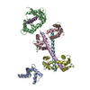 3g43S C: citing same article ( S: Starting model for refinement |
|---|---|
| Similar structure data |

-Links
 PDBj
PDBj


- Assembly
| Deposited unit | 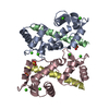
| ||||||||
|---|---|---|---|---|---|---|---|---|---|
| 1 | 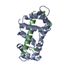
| ||||||||
| 2 | 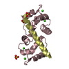
| ||||||||
| Unit cell |
|
-Components
| #1: Protein | Mass: 16950.584 Da / Num. of mol.: 2 / Fragment: UNP residues 1-147 Source method: isolated from a genetically manipulated source Source: (gene. exp.) Homo sapiens (human)Gene: CALM1, CALM, CAM, CAM1, CALM2, CAM2, CAMB, CALM3, CALML2, CAM3, CAMC, CAMIII Production host:  #2: Protein/peptide | Mass: 5658.692 Da / Num. of mol.: 2 / Fragment: UNP residues 828-870 Source method: isolated from a genetically manipulated source Source: (gene. exp.) #3: Chemical | ChemComp-CA /   Mass: 40.078 Da / Num. of mol.: 8 / Source method: obtained synthetically / Formula: Ca Mass: 40.078 Da / Num. of mol.: 8 / Source method: obtained synthetically / Formula: Ca#4: Chemical |   Mass: 96.063 Da / Num. of mol.: 3 / Source method: obtained synthetically / Formula: SO4 Mass: 96.063 Da / Num. of mol.: 3 / Source method: obtained synthetically / Formula: SO4#5: Water | ChemComp-HOH / |  Mass: 18.015 Da / Num. of mol.: 12 / Source method: isolated from a natural source / Formula: H2O Mass: 18.015 Da / Num. of mol.: 12 / Source method: isolated from a natural source / Formula: H2O |
|---|
-Experimental details
-Experiment
| Experiment | Method: X-RAY DIFFRACTION / Number of used crystals: 1 |
|---|
- Sample preparation
| Crystal | Density Matthews: 1.69 Å3/Da / Density % sol: 27.1 % |
|---|---|
| Crystal grow | Temperature: 289 K / Method: vapor diffusion / pH: 6.5 Details: 0.2M ammonium sulfate, 0.1M Bis-Tris (pH 6.5), 25%(w/v) PEG 3,500 |
-Data collection
| Diffraction | Mean temperature: 100 K |
|---|---|
| Diffraction source | Source: SYNCHROTRON / Site: SSRF  / Beamline: BL17U / Wavelength: 0.9791 Å / Beamline: BL17U / Wavelength: 0.9791 Å |
| Detector | Type: ADSC QUANTUM 315r / Detector: CCD / Date: Apr 12, 2014 |
| Radiation | Monochromator: Double crystal / Protocol: SINGLE WAVELENGTH / Monochromatic (M) / Laue (L): M / Scattering type: x-ray |
| Radiation wavelength | Wavelength: 0.9791 Å / Relative weight: 1 |
| Reflection | Resolution: 2.33→50 Å / Num. obs: 12459 / % possible obs: 97.5 % / Redundancy: 2.8 % / Rmerge(I) obs: 0.062 / Net I/σ(I): 23 |
| Reflection shell | Resolution: 2.35→2.39 Å / Redundancy: 2.7 % / Rmerge(I) obs: 0.36 / Mean I/σ(I) obs: 4.1 / Rsym value: 0.36 / % possible all: 98.9 |
- Processing
| Software |
| |||||||||||||||||||||||||||||||||||||||||||||||||||||||||||||||||||||||||||||||||||||||||||||||||||||||||||||||||||||||||||||
|---|---|---|---|---|---|---|---|---|---|---|---|---|---|---|---|---|---|---|---|---|---|---|---|---|---|---|---|---|---|---|---|---|---|---|---|---|---|---|---|---|---|---|---|---|---|---|---|---|---|---|---|---|---|---|---|---|---|---|---|---|---|---|---|---|---|---|---|---|---|---|---|---|---|---|---|---|---|---|---|---|---|---|---|---|---|---|---|---|---|---|---|---|---|---|---|---|---|---|---|---|---|---|---|---|---|---|---|---|---|---|---|---|---|---|---|---|---|---|---|---|---|---|---|---|---|---|
| Refinement | Method to determine structure: MOLECULAR REPLACEMENT Starting model: 3G43 Resolution: 2.33→44.88 Å / Cor.coef. Fo:Fc: 0.926 / Cor.coef. Fo:Fc free: 0.897 / SU B: 25.713 / SU ML: 0.282 / Cross valid method: THROUGHOUT / σ(F): 0 / ESU R: 0.643 / ESU R Free: 0.289 Details: U VALUES : WITH TLS ADDED HYDROGENS HAVE BEEN ADDED IN THE RIDING POSITIONS
| |||||||||||||||||||||||||||||||||||||||||||||||||||||||||||||||||||||||||||||||||||||||||||||||||||||||||||||||||||||||||||||
| Solvent computation | Ion probe radii: 0.8 Å / Shrinkage radii: 0.8 Å / VDW probe radii: 1.2 Å | |||||||||||||||||||||||||||||||||||||||||||||||||||||||||||||||||||||||||||||||||||||||||||||||||||||||||||||||||||||||||||||
| Displacement parameters | Biso max: 93.27 Å2 / Biso mean: 45.433 Å2 / Biso min: 23.49 Å2
| |||||||||||||||||||||||||||||||||||||||||||||||||||||||||||||||||||||||||||||||||||||||||||||||||||||||||||||||||||||||||||||
| Refinement step | Cycle: final / Resolution: 2.33→44.88 Å
| |||||||||||||||||||||||||||||||||||||||||||||||||||||||||||||||||||||||||||||||||||||||||||||||||||||||||||||||||||||||||||||
| Refine LS restraints |
| |||||||||||||||||||||||||||||||||||||||||||||||||||||||||||||||||||||||||||||||||||||||||||||||||||||||||||||||||||||||||||||
| LS refinement shell | Resolution: 2.329→2.389 Å / Total num. of bins used: 20
| |||||||||||||||||||||||||||||||||||||||||||||||||||||||||||||||||||||||||||||||||||||||||||||||||||||||||||||||||||||||||||||
| Refinement TLS params. | Method: refined / Refine-ID: X-RAY DIFFRACTION
| |||||||||||||||||||||||||||||||||||||||||||||||||||||||||||||||||||||||||||||||||||||||||||||||||||||||||||||||||||||||||||||
| Refinement TLS group |
|
