#1: Journal: Infect.Immun. / Year: 2008 Title: An Iron-Binding Protein, Dpr, Decreases Hydrogen Peroxide Stress and Protects Streptococcus Pyogenes Against Multiple Stresses. Authors: Tsou, C. / Chiang-Ni, C. / Lin, Y. / Chuang, W. / Lin, M. / Liu, C. / Wu, J.
History
Deposition
Jun 26, 2009
Deposition site: PDBE / Processing site: PDBE
Revision 1.0
Sep 15, 2009
Provider: repository / Type: Initial release
Revision 1.1
May 8, 2011
Group: Version format compliance
Revision 1.2
Jul 13, 2011
Group: Version format compliance
Revision 1.3
Jul 24, 2019
Group: Data collection / Category: diffrn_source / Item: _diffrn_source.pdbx_synchrotron_site
Monochromator: SI (111), HORIZONTALLY FOCUSSING / Protocol: SINGLE WAVELENGTH / Monochromatic (M) / Laue (L): M / Scattering type: x-ray
Radiation wavelength
Wavelength: 0.8132 Å / Relative weight: 1
Reflection
Resolution: 1.93→43 Å / Num. obs: 20355 / % possible obs: 92.7 % / Observed criterion σ(I): 2 / Redundancy: 7.3 % / Rmerge(I) obs: 0.09 / Net I/σ(I): 16
Reflection shell
Resolution: 1.93→2.03 Å / Redundancy: 6.4 % / Rmerge(I) obs: 0.33 / Mean I/σ(I) obs: 4.9 / % possible all: 79.1
-
Processing
Software
Name
Version
Classification
REFMAC
5.4.0078
refinement
MOSFLM
datareduction
SCALA
datascaling
Refinement
Method to determine structure: OTHER Starting model: NONE Resolution: 1.94→43 Å / Cor.coef. Fo:Fc: 0.963 / Cor.coef. Fo:Fc free: 0.957 / SU B: 1.754 / SU ML: 0.052 / Cross valid method: THROUGHOUT / ESU R: 0.102 / ESU R Free: 0.094 / Stereochemistry target values: MAXIMUM LIKELIHOOD / Details: HYDROGENS HAVE BEEN ADDED IN THE RIDING POSITIONS.
Rfactor
Num. reflection
% reflection
Selection details
Rfree
0.16394
1058
5.2 %
RANDOM
Rwork
0.14565
-
-
-
obs
0.14663
19295
93.91 %
-
Solvent computation
Ion probe radii: 0.8 Å / Shrinkage radii: 0.8 Å / VDW probe radii: 1.2 Å / Solvent model: MASK
 Movie
Movie Controller


 Open data
 Basic information
 Components
 Keywords
 Function and homology information
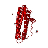
 STREPTOCOCCUS PYOGENES (bacteria)
 X-RAY DIFFRACTION /
  Authors
 Citation
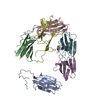
 Structure visualization
 Downloads & links
 Other downloads

 PDBj
PDBj


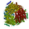
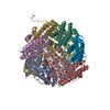
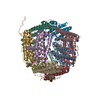
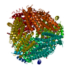
 Assembly

 Mass: 22.990 Da / Num. of mol.: 1 / Source method: obtained synthetically / Formula: Na
 Mass: 55.845 Da / Num. of mol.: 1 / Source method: obtained synthetically / Formula: Fe
 Mass: 92.094 Da / Num. of mol.: 3 / Source method: obtained synthetically / Formula: C3H8O3
 Mass: 18.015 Da / Num. of mol.: 223 / Source method: isolated from a natural source / Formula: H2O
 Sample preparation
 / Beamline: X13 / Wavelength: 0.8132
  Processing