 Movie
Movie Controller

[English] 日本語
 Yorodumi
- PDB-2bw1: Iron-bound crystal structure of Dps-like peroxide resistance prot... -
+ Open data

- Basic information
| Entry | Database: PDB / ID: 2bw1 | ||||||
|---|---|---|---|---|---|---|---|
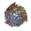
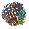
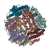
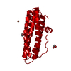
| Title | Iron-bound crystal structure of Dps-like peroxide resistance protein (Dpr) from Streptococcus suis. | ||||||
 Components Components | DPS-LIKE PEROXIDE RESISTANCE PROTEIN | ||||||
 Keywords Keywords | PEROXIDE RESISTANCE / DPR / IRON-BINDING / FERROXIDASE / DPS-FAMILY / FERRITIN-LIKE | ||||||
| Function / homology |  Function and homology information Function and homology informationOxidoreductases; Oxidizing metal ions / ferric iron binding / intracellular iron ion homeostasis / oxidoreductase activity / cytoplasm Similarity search - Function | ||||||
| Biological species |  STREPTOCOCCUS SUIS (bacteria) STREPTOCOCCUS SUIS (bacteria) | ||||||
| Method |  X-RAY DIFFRACTION / SYNCHROTRON / MOLECULAR REPLACEMENT / Resolution: 1.81 Å X-RAY DIFFRACTION / SYNCHROTRON / MOLECULAR REPLACEMENT / Resolution: 1.81 Å | ||||||
 Authors Authors | Kauko, A. / Pulliainen, A. / Haataja, S. / Finne, J. / Papageorgiou, A.C. | ||||||
 Citation Citation | Journal: J. Mol. Biol. / Year: 2006 Title: Iron incorporation in Streptococcus suis Dps-like peroxide resistance protein Dpr requires mobility in the ferroxidase center and leads to the formation of a ferrihydrite-like core. Authors: Kauko, A. / Pulliainen, A.T. / Haataja, S. / Meyer-Klaucke, W. / Finne, J. / Papageorgiou, A.C. | ||||||
| History |
|
- Structure visualization
| Structure viewer | Molecule: MolmilJmol/JSmol |
|---|
- Downloads & links
-Download
| PDBx/mmCIF format | 2bw1.cif.gz | 404.2 KB | Display | PDBx/mmCIF format |
|---|---|---|---|---|
| PDB format | pdb2bw1.ent.gz | 333.2 KB | Display | PDB format |
| PDBx/mmJSON format | 2bw1.json.gz | Tree view | PDBx/mmJSON format | |
| Others |  Other downloads Other downloads |
-Validation report
| Arichive directory | https://data.pdbj.org/pub/pdb/validation_reports/bw/2bw1ftp://data.pdbj.org/pub/pdb/validation_reports/bw/2bw1 | HTTPS FTP |
|---|
-Related structure data


-Links
 PDBj
PDBj


- Assembly
| Deposited unit | 
| ||||||||||||||||||||||||||||||||||||||||||||||||
|---|---|---|---|---|---|---|---|---|---|---|---|---|---|---|---|---|---|---|---|---|---|---|---|---|---|---|---|---|---|---|---|---|---|---|---|---|---|---|---|---|---|---|---|---|---|---|---|---|---|
| 1 |
| ||||||||||||||||||||||||||||||||||||||||||||||||
| Unit cell |
| ||||||||||||||||||||||||||||||||||||||||||||||||
| Noncrystallographic symmetry (NCS) | NCS oper:
|
-Components
| #1: Protein | Mass: 18569.938 Da / Num. of mol.: 12 / Mutation: YES Source method: isolated from a genetically manipulated source Details: N-TERMINUS TRUNCATED AND FIRST SEVEN RESIDUES REMOVED. Source: (gene. exp.) STREPTOCOCCUS SUIS (bacteria) / Strain: D282 / Plasmid: PET30EK / Production host: #2: Chemical | ChemComp-EPE /   Mass: 238.305 Da / Num. of mol.: 4 / Source method: obtained synthetically / Formula: C8H18N2O4S / Comment: pH buffer*YM Mass: 238.305 Da / Num. of mol.: 4 / Source method: obtained synthetically / Formula: C8H18N2O4S / Comment: pH buffer*YM#3: Chemical | ChemComp-FE /   Mass: 55.845 Da / Num. of mol.: 12 / Source method: obtained synthetically / Formula: Fe Mass: 55.845 Da / Num. of mol.: 12 / Source method: obtained synthetically / Formula: Fe#4: Chemical | ChemComp-CA / |   Mass: 40.078 Da / Num. of mol.: 1 / Source method: obtained synthetically / Formula: Ca Mass: 40.078 Da / Num. of mol.: 1 / Source method: obtained synthetically / Formula: Ca#5: Water | ChemComp-HOH / |  Mass: 18.015 Da / Num. of mol.: 1502 / Source method: isolated from a natural source / Formula: H2O Mass: 18.015 Da / Num. of mol.: 1502 / Source method: isolated from a natural source / Formula: H2OCompound details | ENGINEERED MUTATION GLN 8 TO GLY IN CHAINS A-L. ONLY THE CHAIN G MUTATION WAS VISIBLE IN THE ...ENGINEERED | Sequence details | UNIPROT ENTRY HAS FULL LENGTH PROTEIN. PROTEIN DESCRIBED IN THIS PDB-ENTRY HAS TRUNCATED N-TERMINUS ...UNIPROT ENTRY HAS FULL LENGTH PROTEIN. PROTEIN DESCRIBED IN THIS PDB-ENTRY HAS TRUNCATED N-TERMINUS WITH FIRST 7 RESIDUES MISSING AND Q8G MUTATION. | |
|---|
-Experimental details
-Experiment
| Experiment | Method: X-RAY DIFFRACTION / Number of used crystals: 1 |
|---|
- Sample preparation
| Crystal | Density Matthews: 2.3 Å3/Da / Density % sol: 46.6 % Description: A 2.81A DATASET FOR THIS STRUCTURE WAS USED AS STARTING MODEL. |
|---|---|
| Crystal grow | Temperature: 289 K / Method: vapor diffusion, hanging drop / pH: 7.4 Details: 2UL AND 2UL VOLUME DROP, 30 % PEG 400, 0.2 M CACL2, 0.1 M HEPES-NAOH, PH 7.4, HANGING DROP, 16C |
-Data collection
| Diffraction | Mean temperature: 100 K |
|---|---|
| Diffraction source | Source: SYNCHROTRON / Site: EMBL/DESY, HAMBURG  / Beamline: X11 / Wavelength: 0.8128 / Beamline: X11 / Wavelength: 0.8128 |
| Detector | Type: MARRESEARCH / Detector: CCD / Date: Nov 19, 2004 / Details: MIRRORS |
| Radiation | Monochromator: TRIANGULAR HORIZONTAL- FOCUSING SI III / Protocol: SINGLE WAVELENGTH / Monochromatic (M) / Laue (L): M / Scattering type: x-ray |
| Radiation wavelength | Wavelength: 0.8128 Å / Relative weight: 1 |
| Reflection | Resolution: 1.8→20 Å / Num. obs: 188319 / % possible obs: 98 % / Observed criterion σ(I): -3 / Redundancy: 8 % / Biso Wilson estimate: 30.9 Å2 / Rmerge(I) obs: 0.05 / Rsym value: 0.06 / Net I/σ(I): 21 |
| Reflection shell | Resolution: 1.8→1.91 Å / Redundancy: 7.2 % / Rmerge(I) obs: 0.24 / Mean I/σ(I) obs: 6 / Rsym value: 0.34 / % possible all: 91 |
- Processing
| Software |
| ||||||||||||||||||||||||||||||||||||||||||||||||||||||||||||||||||||||||||||||||||||||||||||||||||||||||||||||||||||||||||||||||||||||||||||||||||||||||||||||||||||||||||||||||||||||
|---|---|---|---|---|---|---|---|---|---|---|---|---|---|---|---|---|---|---|---|---|---|---|---|---|---|---|---|---|---|---|---|---|---|---|---|---|---|---|---|---|---|---|---|---|---|---|---|---|---|---|---|---|---|---|---|---|---|---|---|---|---|---|---|---|---|---|---|---|---|---|---|---|---|---|---|---|---|---|---|---|---|---|---|---|---|---|---|---|---|---|---|---|---|---|---|---|---|---|---|---|---|---|---|---|---|---|---|---|---|---|---|---|---|---|---|---|---|---|---|---|---|---|---|---|---|---|---|---|---|---|---|---|---|---|---|---|---|---|---|---|---|---|---|---|---|---|---|---|---|---|---|---|---|---|---|---|---|---|---|---|---|---|---|---|---|---|---|---|---|---|---|---|---|---|---|---|---|---|---|---|---|---|---|
| Refinement | Method to determine structure: MOLECULAR REPLACEMENT Starting model: STRUCTURE FROM PREVIOUS DATASET Resolution: 1.81→19.53 Å / Cor.coef. Fo:Fc: 0.956 / Cor.coef. Fo:Fc free: 0.937 / SU B: 2.178 / SU ML: 0.069 / Cross valid method: THROUGHOUT / ESU R: 0.125 / ESU R Free: 0.129 / Stereochemistry target values: MAXIMUM LIKELIHOOD Details: LAST CYCLE OF REFINEMENT WAS DONE WITHOUT R-FREE SET. STATISTICS RELATED TO R-FREE ARE FORM SECOND LAST CYCLE. CRYSTAL SOAKED FOR 10 MIN IN 10 MM (NH4) 2FE(SO4)2 AND 2.5 PERCENT (SO2NA)2 I.E. REDUCTANT.
| ||||||||||||||||||||||||||||||||||||||||||||||||||||||||||||||||||||||||||||||||||||||||||||||||||||||||||||||||||||||||||||||||||||||||||||||||||||||||||||||||||||||||||||||||||||||
| Solvent computation | Ion probe radii: 0.8 Å / Shrinkage radii: 0.8 Å / VDW probe radii: 1.2 Å / Solvent model: MASK | ||||||||||||||||||||||||||||||||||||||||||||||||||||||||||||||||||||||||||||||||||||||||||||||||||||||||||||||||||||||||||||||||||||||||||||||||||||||||||||||||||||||||||||||||||||||
| Displacement parameters | Biso mean: 24.4 Å2 | ||||||||||||||||||||||||||||||||||||||||||||||||||||||||||||||||||||||||||||||||||||||||||||||||||||||||||||||||||||||||||||||||||||||||||||||||||||||||||||||||||||||||||||||||||||||
| Refinement step | Cycle: LAST / Resolution: 1.81→19.53 Å
| ||||||||||||||||||||||||||||||||||||||||||||||||||||||||||||||||||||||||||||||||||||||||||||||||||||||||||||||||||||||||||||||||||||||||||||||||||||||||||||||||||||||||||||||||||||||
| Refine LS restraints |
|