 Movie
Movie Controller

[English] 日本語
 Yorodumi
- PDB-1umn: Crystal structure of Dps-like peroxide resistance protein (Dpr) f... -
+ Open data

- Basic information
| Entry | Database: PDB / ID: 1umn | ||||||
|---|---|---|---|---|---|---|---|
| Title | Crystal structure of Dps-like peroxide resistance protein (Dpr) from Streptococcus suis | ||||||
 Components Components | DPS-LIKE PEROXIDE RESISTANCE PROTEIN | ||||||
 Keywords Keywords | PEROXIDE RESISTANCE / IRON-BINDING / FERROXIDASE / DPS-FAMILY / FERRITIN-LIKE | ||||||
| Function / homology |  Function and homology information Function and homology informationOxidoreductases; Oxidizing metal ions / ferric iron binding / intracellular iron ion homeostasis / oxidoreductase activity / cytoplasm Similarity search - Function | ||||||
| Biological species |  STREPTOCOCCUS SUIS (bacteria) STREPTOCOCCUS SUIS (bacteria) | ||||||
| Method |  X-RAY DIFFRACTION / SYNCHROTRON / MOLECULAR REPLACEMENT / Resolution: 1.95 Å X-RAY DIFFRACTION / SYNCHROTRON / MOLECULAR REPLACEMENT / Resolution: 1.95 Å | ||||||
 Authors Authors | Kauko, A. / Haataja, S. / Pulliainen, A. / Finne, J. / Papageorgiou, A.C. | ||||||
 Citation Citation | Journal: J. Mol. Biol. / Year: 2004 Title: Crystal structure of Streptococcus suis Dps-like peroxide resistance protein Dpr: implications for iron incorporation. Authors: Kauko, A. / Haataja, S. / Pulliainen, A.T. / Finne, J. / Papageorgiou, A.C. | ||||||
| History |
|
- Structure visualization
| Structure viewer | Molecule: MolmilJmol/JSmol |
|---|
- Downloads & links
-Download
| PDBx/mmCIF format | 1umn.cif.gz | 401.8 KB | Display | PDBx/mmCIF format |
|---|---|---|---|---|
| PDB format | pdb1umn.ent.gz | 329.8 KB | Display | PDB format |
| PDBx/mmJSON format | 1umn.json.gz | Tree view | PDBx/mmJSON format | |
| Others |  Other downloads Other downloads |
-Validation report
| Arichive directory | https://data.pdbj.org/pub/pdb/validation_reports/um/1umnftp://data.pdbj.org/pub/pdb/validation_reports/um/1umn | HTTPS FTP |
|---|
-Related structure data
| Related structure data |  1qghS S: Starting model for refinement |
|---|---|
| Similar structure data |

-Links
 PDBj
PDBj


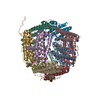
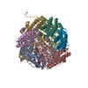
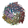
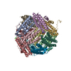
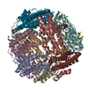
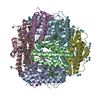
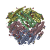
- Assembly
| Deposited unit | 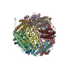
| ||||||||||||||||||||||||||||||||||||||||||||||||||||
|---|---|---|---|---|---|---|---|---|---|---|---|---|---|---|---|---|---|---|---|---|---|---|---|---|---|---|---|---|---|---|---|---|---|---|---|---|---|---|---|---|---|---|---|---|---|---|---|---|---|---|---|---|---|
| 1 |
| ||||||||||||||||||||||||||||||||||||||||||||||||||||
| Unit cell |
| ||||||||||||||||||||||||||||||||||||||||||||||||||||
| Noncrystallographic symmetry (NCS) | NCS oper:
|
-Components
| #1: Protein | Mass: 18641.014 Da / Num. of mol.: 12 Source method: isolated from a genetically manipulated source Details: N-TERMINUS TRUNCATED FIRST SEVEN RESIDUES REMOVED. / Source: (gene. exp.) STREPTOCOCCUS SUIS (bacteria) / Strain: D282 / Plasmid: PET30EK / Production host: #2: Chemical | ChemComp-CA /   Mass: 40.078 Da / Num. of mol.: 16 / Source method: obtained synthetically / Formula: Ca Mass: 40.078 Da / Num. of mol.: 16 / Source method: obtained synthetically / Formula: Ca#3: Chemical |   Mass: 35.453 Da / Num. of mol.: 3 / Source method: obtained synthetically / Formula: Cl Mass: 35.453 Da / Num. of mol.: 3 / Source method: obtained synthetically / Formula: Cl#4: Chemical | ChemComp-EPE /   Mass: 238.305 Da / Num. of mol.: 4 / Source method: obtained synthetically / Formula: C8H18N2O4S / Comment: pH buffer*YM Mass: 238.305 Da / Num. of mol.: 4 / Source method: obtained synthetically / Formula: C8H18N2O4S / Comment: pH buffer*YM#5: Water | ChemComp-HOH / |  Mass: 18.015 Da / Num. of mol.: 1685 / Source method: isolated from a natural source / Formula: H2O Mass: 18.015 Da / Num. of mol.: 1685 / Source method: isolated from a natural source / Formula: H2O |
|---|
-Experimental details
-Experiment
| Experiment | Method: X-RAY DIFFRACTION / Number of used crystals: 1 |
|---|
- Sample preparation
| Crystal | Density Matthews: 2.3 Å3/Da / Density % sol: 44 % |
|---|---|
| Crystal grow | Temperature: 289 K / Method: vapor diffusion, hanging drop / pH: 7.4 Details: HANGING DROP METHOD 2.0 UL 10 MG/ML PROTEIN + 2.0 UL RESERVOUR SOLUTION 27 % PEG 400, 0.2 M CACL2, 0.1M HEPES-NAOH, PH 7.4 +16C FEW DAYS |
-Data collection
| Diffraction | Mean temperature: 100 K |
|---|---|
| Diffraction source | Source: SYNCHROTRON / Site: ELETTRA  / Beamline: 5.2R / Wavelength: 1 / Beamline: 5.2R / Wavelength: 1 |
| Detector | Type: MARRESEARCH / Detector: CCD / Date: Feb 15, 2002 / Details: MIRRORS |
| Radiation | Monochromator: SI CRYSTAL / Protocol: SINGLE WAVELENGTH / Monochromatic (M) / Laue (L): M / Scattering type: x-ray |
| Radiation wavelength | Wavelength: 1 Å / Relative weight: 1 |
| Reflection | Resolution: 1.95→20 Å / Num. obs: 146502 / % possible obs: 97.7 % / Observed criterion σ(I): -3 / Redundancy: 6.3 % / Biso Wilson estimate: 15 Å2 / Rmerge(I) obs: 0.056 / Net I/σ(I): 12 |
| Reflection shell | Resolution: 1.95→2.01 Å / Redundancy: 4.9 % / Rmerge(I) obs: 0.272 / Mean I/σ(I) obs: 1.7 / % possible all: 91.3 |
- Processing
| Software |
| ||||||||||||||||||||||||||||||||||||||||||||||||||||||||||||
|---|---|---|---|---|---|---|---|---|---|---|---|---|---|---|---|---|---|---|---|---|---|---|---|---|---|---|---|---|---|---|---|---|---|---|---|---|---|---|---|---|---|---|---|---|---|---|---|---|---|---|---|---|---|---|---|---|---|---|---|---|---|
| Refinement | Method to determine structure: MOLECULAR REPLACEMENT Starting model: PDB ENTRY 1QGH Resolution: 1.95→18.08 Å / Rfactor Rfree error: 0.003 / Data cutoff high absF: 3678334.77 / Isotropic thermal model: RESTRAINED / Cross valid method: THROUGHOUT / σ(F): 0
| ||||||||||||||||||||||||||||||||||||||||||||||||||||||||||||
| Solvent computation | Solvent model: FLAT MODEL / Bsol: 53.3696 Å2 / ksol: 0.377455 e/Å3 | ||||||||||||||||||||||||||||||||||||||||||||||||||||||||||||
| Displacement parameters | Biso mean: 22.3 Å2
| ||||||||||||||||||||||||||||||||||||||||||||||||||||||||||||
| Refine analyze |
| ||||||||||||||||||||||||||||||||||||||||||||||||||||||||||||
| Refinement step | Cycle: LAST / Resolution: 1.95→18.08 Å
| ||||||||||||||||||||||||||||||||||||||||||||||||||||||||||||
| Refine LS restraints |
| ||||||||||||||||||||||||||||||||||||||||||||||||||||||||||||
| LS refinement shell | Resolution: 1.95→2.07 Å / Rfactor Rfree error: 0.008 / Total num. of bins used: 6
| ||||||||||||||||||||||||||||||||||||||||||||||||||||||||||||
| Xplor file |
|
