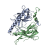
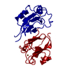
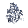
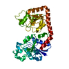
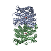
PDB-9rdu: Crystal structure of the Protein-Kinase A catalytic subunit from Cricetulus griseus in complex with F001 Method: X-RAY DIFFRACTION / Resolution: 1.42 Å
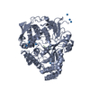
PDB-9rdv: Crystal Structure of the Protein-Kinase A catalytic subunit from Cricetulus griseus in complex with F005 Method: X-RAY DIFFRACTION / Resolution: 1.32 Å
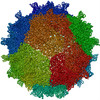
PDB-9rdw: Crystal Structure of the Protein-Kinase A catalytic subunit from Cricetulus griseus in complex with F009 Method: X-RAY DIFFRACTION / Resolution: 1.44 Å
PDB-9rdx: Crystal Structure of the Protein-Kinase A catalytic subunit from Cricetulus griseus in complex with F012 Method: X-RAY DIFFRACTION / Resolution: 1.5 Å
PDB-9rdy: Crystal Structure of the Protein-Kinase A catalytic subunit from Cricetulus griseus in complex with F024 Method: X-RAY DIFFRACTION / Resolution: 1.5 Å
PDB-9rdz: Crystal Structure of the Protein-Kinase A catalytic subunit from Cricetulus griseus in complex with F030 Method: X-RAY DIFFRACTION / Resolution: 1.64 Å
PDB-9re0: Crystal Structure of the Protein-Kinase A catalytic subunit from Cricetulus griseus in complex with F032 Method: X-RAY DIFFRACTION / Resolution: 1.58 Å
PDB-9re1: Crystal Structure of the Protein-Kinase A catalytic subunit from Cricetulus griseus in complex with F055 Method: X-RAY DIFFRACTION / Resolution: 1.41 Å
PDB-9re2: Crystal Structure of the Protein-Kinase A catalytic subunit from Cricetulus griseus in complex with F058 Method: X-RAY DIFFRACTION / Resolution: 1.68 Å
PDB-9re3: Crystal Structure of the Protein-Kinase A catalytic subunit from Cricetulus griseus in complex with F070 Method: X-RAY DIFFRACTION / Resolution: 1.57 Å
PDB-9re4: Crystal Structure of the Protein-Kinase A catalytic subunit from Cricetulus griseus in complex with F073 Method: X-RAY DIFFRACTION / Resolution: 1.47 Å
PDB-9re5: Crystal Structure of the Protein-Kinase A catalytic subunit from Cricetulus griseus in complex with F074 Method: X-RAY DIFFRACTION / Resolution: 1.7 Å
PDB-9re6: Crystal Structure of the Protein-Kinase A catalytic subunit from Cricetulus griseus in complex with F102 Method: X-RAY DIFFRACTION / Resolution: 1.45 Å
PDB-9re7: Crystal Structure of the Protein-Kinase A catalytic subunit from Cricetulus griseus in complex with F134 Method: X-RAY DIFFRACTION / Resolution: 1.46 Å
PDB-9re8: Crystal Structure of the Protein-Kinase A catalytic subunit from Cricetulus griseus in complex with F138 Method: X-RAY DIFFRACTION / Resolution: 1.5 Å
PDB-9re9: Crystal Structure of the Protein-Kinase A catalytic subunit from Cricetulus griseus in complex with F145 Method: X-RAY DIFFRACTION / Resolution: 1.41 Å
PDB-9rea: Crystal Structure of the Protein-Kinase A catalytic subunit from Cricetulus griseus in complex with F168 Method: X-RAY DIFFRACTION / Resolution: 1.52 Å
PDB-9reb: Crystal Structure of the Protein-Kinase A catalytic subunit from Cricetulus griseus in complex with F184 Method: X-RAY DIFFRACTION / Resolution: 1.39 Å
PDB-9rec: Crystal Structure of the Protein-Kinase A catalytic subunit from Cricetulus griseus in complex with F186 Method: X-RAY DIFFRACTION / Resolution: 1.2 Å
PDB-9red: Crystal Structure of the Protein-Kinase A catalytic subunit from Cricetulus griseus in complex with F188 Method: X-RAY DIFFRACTION / Resolution: 1.31 Å
PDB-9ree: Crystal Structure of the Protein-Kinase A catalytic subunit from Cricetulus griseus in complex with F189 Method: X-RAY DIFFRACTION / Resolution: 1.34 Å
PDB-9ref: Crystal Structure of the Protein-Kinase A catalytic subunit from Cricetulus griseus in complex with F203 Method: X-RAY DIFFRACTION / Resolution: 1.42 Å
PDB-9reg: Crystal Structure of the Protein-Kinase A catalytic subunit from Cricetulus griseus in complex with F225 Method: X-RAY DIFFRACTION / Resolution: 1.35 Å
PDB-9reh: Crystal Structure of the Protein-Kinase A catalytic subunit from Cricetulus griseus in complex with F236 Method: X-RAY DIFFRACTION / Resolution: 1.33 Å
PDB-9rei: Crystal Structure of the Protein-Kinase A catalytic subunit from Cricetulus griseus in complex with F248 Method: X-RAY DIFFRACTION / Resolution: 1.42 Å
PDB-9rej: Crystal Structure of the Protein-Kinase A catalytic subunit from Cricetulus griseus in complex with F264 Method: X-RAY DIFFRACTION / Resolution: 1.4 Å
PDB-9rek: Crystal Structure of the Protein-Kinase A catalytic subunit from Cricetulus griseus in complex with F274 Method: X-RAY DIFFRACTION / Resolution: 1.38 Å
PDB-9rel: Crystal Structure of the Protein-Kinase A catalytic subunit from Cricetulus griseus in complex with F283 Method: X-RAY DIFFRACTION / Resolution: 1.42 Å
PDB-9rem: Crystal Structure of the Protein-Kinase A catalytic subunit from Cricetulus griseus in complex with F294 Method: X-RAY DIFFRACTION / Resolution: 1.43 Å
PDB-9ren: Crystal Structure of the Protein-Kinase A catalytic subunit from Cricetulus griseus in complex with F296 Method: X-RAY DIFFRACTION / Resolution: 1.38 Å
PDB-9reo: Crystal Structure of the Protein-Kinase A catalytic subunit from Cricetulus griseus in complex with F299 Method: X-RAY DIFFRACTION / Resolution: 1.45 Å
PDB-9rep: Crystal Structure of the Protein-Kinase A catalytic subunit from Cricetulus griseus in complex with F304 Method: X-RAY DIFFRACTION / Resolution: 1.44 Å
PDB-9req: Crystal Structure of the Protein-Kinase A catalytic subunit from Cricetulus griseus in complex with F310 Method: X-RAY DIFFRACTION / Resolution: 1.5 Å
PDB-9rer: Crystal Structure of the Protein-Kinase A catalytic subunit from Cricetulus griseus in complex with F312 Method: X-RAY DIFFRACTION / Resolution: 1.45 Å
PDB-9res: Crystal Structure of the Protein-Kinase A catalytic subunit from Cricetulus griseus in complex with F313 Method: X-RAY DIFFRACTION / Resolution: 1.5 Å
PDB-9ret: Crystal Structure of the Protein-Kinase A catalytic subunit from Cricetulus griseus in complex with F322 Method: X-RAY DIFFRACTION / Resolution: 1.75 Å
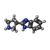
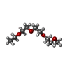
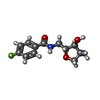
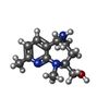
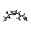
Chemicals
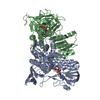
PDB-1je8: Two-Component response regulator NarL/DNA Complex: DNA Bending Found in a High Affinity Site
PDB-1jep: Chalcone Isomerase Complexed with 4'-hydroxyflavanone
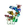
PDB-1jeo: Crystal Structure of the Hypothetical Protein MJ1247 from Methanococcus jannaschii at 2.0 A Resolution Infers a Molecular Function of 3-Hexulose-6-Phosphate isomerase.
PDB-1jen: HUMAN S-ADENOSYLMETHIONINE DECARBOXYLASE
PDB-1jem: NMR STRUCTURE OF HISTIDINE PHOSPHORYLATED FORM OF THE PHOSPHOCARRIER HISTIDINE CONTAINING PROTEIN FROM BACILLUS SUBTILIS, NMR, 25 STRUCTURES
In the structure databanks used in Yorodumi, some data are registered as the other names, "COVID-19 virus" and "2019-nCoV". Here are the details of the virus and the list of structure data.
Jan 31, 2019. EMDB accession codes are about to change! (news from PDBe EMDB page)
EMDB accession codes are about to change! (news from PDBe EMDB page)
The allocation of 4 digits for EMDB accession codes will soon come to an end. Whilst these codes will remain in use, new EMDB accession codes will include an additional digit and will expand incrementally as the available range of codes is exhausted. The current 4-digit format prefixed with “EMD-” (i.e. EMD-XXXX) will advance to a 5-digit format (i.e. EMD-XXXXX), and so on. It is currently estimated that the 4-digit codes will be depleted around Spring 2019, at which point the 5-digit format will come into force.
The EM Navigator/Yorodumi systems omit the EMD- prefix.
Related info.:Q: What is EMD? / ID/Accession-code notation in Yorodumi/EM Navigator
 Movie
Movie Controller
 Structure viewers
 About Yorodumi Papers


 Authors
 External links


 Keywords
 cricetulus griseus (Chinese hamster)