 Movie
Movie Controller

[English] 日本語
 Yorodumi
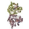
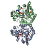
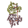
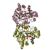
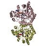
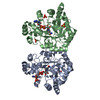
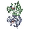
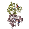
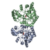
- PDB-1jez: THE STRUCTURE OF XYLOSE REDUCTASE, A DIMERIC ALDO-KETO REDUCTASE ... -
+ Open data

- Basic information
| Entry | Database: PDB / ID: 1jez | ||||||
|---|---|---|---|---|---|---|---|
| Title | THE STRUCTURE OF XYLOSE REDUCTASE, A DIMERIC ALDO-KETO REDUCTASE FROM CANDIDA TENUIS | ||||||
 Components Components | XYLOSE REDUCTASE | ||||||
 Keywords Keywords | OXIDOREDUCTASE / TIM BARREL / ALDO-KETO REDUCTASE / NADPH / NADH | ||||||
| Function / homology |  Function and homology information Function and homology informationD-xylose reductase [NAD(P)H] / D-xylose reductase (NADPH) activity / D-xylose catabolic process Similarity search - Function | ||||||
| Biological species |  Candida tenuis (fungus) Candida tenuis (fungus) | ||||||
| Method |  X-RAY DIFFRACTION / MOLECULAR REPLACEMENT / Resolution: 2.2 Å X-RAY DIFFRACTION / MOLECULAR REPLACEMENT / Resolution: 2.2 Å | ||||||
 Authors Authors | Kavanagh, K.L. / Klimacek, M. / Nidetzky, B. / Wilson, D.K. | ||||||
 Citation Citation | Journal: Biochemistry / Year: 2002 Title: The structure of apo and holo forms of xylose reductase, a dimeric aldo-keto reductase from Candida tenuis. Authors: Kavanagh, K.L. / Klimacek, M. / Nidetzky, B. / Wilson, D.K. | ||||||
| History |
|
- Structure visualization
| Structure viewer | Molecule: MolmilJmol/JSmol |
|---|
- Downloads & links
-Download
| PDBx/mmCIF format | 1jez.cif.gz | 136.1 KB | Display | PDBx/mmCIF format |
|---|---|---|---|---|
| PDB format | pdb1jez.ent.gz | 106.4 KB | Display | PDB format |
| PDBx/mmJSON format | 1jez.json.gz | Tree view | PDBx/mmJSON format | |
| Others |  Other downloads Other downloads |
-Validation report
| Arichive directory | https://data.pdbj.org/pub/pdb/validation_reports/je/1jezftp://data.pdbj.org/pub/pdb/validation_reports/je/1jez | HTTPS FTP |
|---|
-Related structure data
| Related structure data | 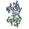 1k8cC 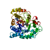 1adsS C: citing same article ( S: Starting model for refinement |
|---|---|
| Similar structure data |

-Links
 PDBj
PDBj
- Assembly
| Deposited unit | 
| ||||||||||
|---|---|---|---|---|---|---|---|---|---|---|---|
| 1 |
| ||||||||||
| Unit cell |
|
-Components
| #1: Protein | Mass: 36062.199 Da / Num. of mol.: 2 Source method: isolated from a genetically manipulated source Source: (gene. exp.) Candida tenuis (fungus) / Gene: XYLR / Plasmid: PBEACT.LI / Species (production host): Escherichia coli / Production host:  #2: Water | ChemComp-HOH / |  Mass: 18.015 Da / Num. of mol.: 407 / Source method: isolated from a natural source / Formula: H2O Mass: 18.015 Da / Num. of mol.: 407 / Source method: isolated from a natural source / Formula: H2O |
|---|
-Experimental details
-Experiment
| Experiment | Method: X-RAY DIFFRACTION / Number of used crystals: 1 |
|---|
- Sample preparation
| Crystal | Density Matthews: 2.34 Å3/Da / Density % sol: 47.4 % | ||||||||||||||||||||||||||||||||||||||||||||||||
|---|---|---|---|---|---|---|---|---|---|---|---|---|---|---|---|---|---|---|---|---|---|---|---|---|---|---|---|---|---|---|---|---|---|---|---|---|---|---|---|---|---|---|---|---|---|---|---|---|---|
| Crystal grow | Temperature: 298 K / Method: vapor diffusion, hanging drop / pH: 5.6 Details: 30% PEG 4000, 200mM ammonium acetate, 100 mM sodium citrate, pH 5.6, VAPOR DIFFUSION, HANGING DROP at 298K | ||||||||||||||||||||||||||||||||||||||||||||||||
| Crystal grow | *PLUS | ||||||||||||||||||||||||||||||||||||||||||||||||
| Components of the solutions | *PLUS
|
-Data collection
| Diffraction | Mean temperature: 100 K |
|---|---|
| Diffraction source | Source: ROTATING ANODE / Type: RIGAKU / Wavelength: 1.5418 Å |
| Detector | Type: RIGAKU RAXIS IV / Detector: IMAGE PLATE / Date: Feb 16, 2001 / Details: mirrors |
| Radiation | Monochromator: Ni MIRROR + Ni FILTER / Protocol: SINGLE WAVELENGTH / Monochromatic (M) / Laue (L): M / Scattering type: x-ray |
| Radiation wavelength | Wavelength: 1.5418 Å / Relative weight: 1 |
| Reflection | Resolution: 2.2→100 Å / Num. all: 33863 / Num. obs: 33457 / % possible obs: 98.8 % / Observed criterion σ(F): 0 / Observed criterion σ(I): 0 / Redundancy: 3.26 % / Biso Wilson estimate: 15.9 Å2 / Rmerge(I) obs: 0.089 / Net I/σ(I): 12.8 |
| Reflection shell | Resolution: 2.2→2.28 Å / Redundancy: 3.1 % / Rmerge(I) obs: 0.406 / Mean I/σ(I) obs: 3.1 / % possible all: 97.3 |
| Reflection | *PLUS Lowest resolution: 100 Å / Num. measured all: 109237 / Rmerge(I) obs: 0.089 |
| Reflection shell | *PLUS % possible obs: 97.3 % / Rmerge(I) obs: 0.406 |
- Processing
| Software |
| ||||||||||||||||||||||||||||||||||||
|---|---|---|---|---|---|---|---|---|---|---|---|---|---|---|---|---|---|---|---|---|---|---|---|---|---|---|---|---|---|---|---|---|---|---|---|---|---|
| Refinement | Method to determine structure: MOLECULAR REPLACEMENT Starting model: modified 1ADS Resolution: 2.2→20 Å / Cross valid method: THROUGHOUT / σ(F): 0 / σ(I): 0 / Stereochemistry target values: Engh & Huber
| ||||||||||||||||||||||||||||||||||||
| Displacement parameters |
| ||||||||||||||||||||||||||||||||||||
| Refinement step | Cycle: LAST / Resolution: 2.2→20 Å
| ||||||||||||||||||||||||||||||||||||
| Refine LS restraints |
| ||||||||||||||||||||||||||||||||||||
| Refinement | *PLUS Lowest resolution: 20 Å / Num. reflection obs: 30391 / Rfactor obs: 0.179 / Rfactor Rfree: 0.233 / Rfactor Rwork: 0.179 | ||||||||||||||||||||||||||||||||||||
| Solvent computation | *PLUS | ||||||||||||||||||||||||||||||||||||
| Displacement parameters | *PLUS | ||||||||||||||||||||||||||||||||||||
| Refine LS restraints | *PLUS
|
