 Movie
Movie Controller

[English] 日本語
 Yorodumi
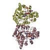
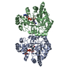
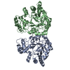
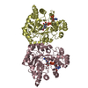
- PDB-1z9a: Crystal Structure Of The Asn-309 To Asp Mutant Of Candida Tenuis ... -
+ Open data

- Basic information
| Entry | Database: PDB / ID: 1z9a | ||||||
|---|---|---|---|---|---|---|---|
| Title | Crystal Structure Of The Asn-309 To Asp Mutant Of Candida Tenuis Xylose Reductase (Akr2B5) Bound To Nad+ | ||||||
 Components Components | NAD(P)H-dependent D-xylose reductase | ||||||
 Keywords Keywords | OXIDOREDUCTASE / beta-alpha-barrel / AKR / aldo-keto reductase / xylose reductase / candida tenuis / substrate selectivity / ketone reduction / structure-activity correlation | ||||||
| Function / homology |  Function and homology information Function and homology informationD-xylose reductase [NAD(P)H] / D-xylose reductase (NADPH) activity / D-xylose catabolic process Similarity search - Function | ||||||
| Biological species |  Candida tenuis (fungus) Candida tenuis (fungus) | ||||||
| Method |  X-RAY DIFFRACTION / isomorphous / Resolution: 2.4 Å X-RAY DIFFRACTION / isomorphous / Resolution: 2.4 Å | ||||||
 Authors Authors | Kratzer, R. / Leitgeb, S. / Wilson, D.K. / Nidetzky, B. | ||||||
 Citation Citation | Journal: Biochem.J. / Year: 2006 Title: Probing the substrate binding site of Candida tenuis xylose reductase (AKR2B5) with site-directed mutagenesis Authors: Kratzer, R. / Leitgeb, S. / Wilson, D.K. / Nidetzky, B. | ||||||
| History |
|
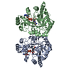
- Structure visualization
| Structure viewer | Molecule: MolmilJmol/JSmol |
|---|
- Downloads & links
-Download
| PDBx/mmCIF format | 1z9a.cif.gz | 277.4 KB | Display | PDBx/mmCIF format |
|---|---|---|---|---|
| PDB format | pdb1z9a.ent.gz | 224.2 KB | Display | PDB format |
| PDBx/mmJSON format | 1z9a.json.gz | Tree view | PDBx/mmJSON format | |
| Others |  Other downloads Other downloads |
-Validation report
| Arichive directory | https://data.pdbj.org/pub/pdb/validation_reports/z9/1z9aftp://data.pdbj.org/pub/pdb/validation_reports/z9/1z9a | HTTPS FTP |
|---|
-Related structure data
| Similar structure data |
|---|
-Links
 PDBj
PDBj
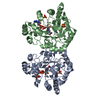
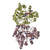
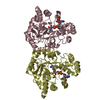
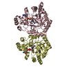
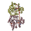
- Assembly
| Deposited unit | 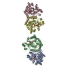
| ||||||||||||
|---|---|---|---|---|---|---|---|---|---|---|---|---|---|
| 1 | 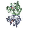
| ||||||||||||
| 2 | 
| ||||||||||||
| 3 | 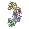
| ||||||||||||
| Unit cell |
| ||||||||||||
| Components on special symmetry positions |
|
-Components
| #1: Protein | Mass: 35931.992 Da / Num. of mol.: 4 / Mutation: N310D Source method: isolated from a genetically manipulated source Source: (gene. exp.) Candida tenuis (fungus) / Gene: XYL1, XYLR / Plasmid: pET11 / Species (production host): Escherichia coli / Production host:  References: UniProt: O74237, Oxidoreductases; Acting on the CH-OH group of donors; With NAD+ or NADP+ as acceptor #2: Chemical | ChemComp-NAD /   Mass: 663.425 Da / Num. of mol.: 4 / Source method: obtained synthetically / Formula: C21H27N7O14P2 / Comment: NAD*YM Mass: 663.425 Da / Num. of mol.: 4 / Source method: obtained synthetically / Formula: C21H27N7O14P2 / Comment: NAD*YM#3: Water | ChemComp-HOH / |  Mass: 18.015 Da / Num. of mol.: 789 / Source method: isolated from a natural source / Formula: H2O Mass: 18.015 Da / Num. of mol.: 789 / Source method: isolated from a natural source / Formula: H2O |
|---|
-Experimental details
-Experiment
| Experiment | Method: X-RAY DIFFRACTION / Number of used crystals: 1 |
|---|
- Sample preparation
| Crystal | Density Matthews: 3.23 Å3/Da / Density % sol: 61.87 % |
|---|---|
| Crystal grow | Temperature: 298 K / Method: vapor diffusion, hanging drop / pH: 6.4 Details: ammonium sulfate, sodium acetate, sodium citrate, pH 6.4, VAPOR DIFFUSION, HANGING DROP, temperature 298K |
-Data collection
| Diffraction | Mean temperature: 100 K |
|---|---|
| Diffraction source | Source: ROTATING ANODE / Type: RIGAKU RU300 / Wavelength: 1.5418 Å |
| Detector | Type: RIGAKU RAXIS IV / Detector: IMAGE PLATE / Date: Jan 28, 2004 |
| Radiation | Monochromator: YALE MIRRORS / Protocol: SINGLE WAVELENGTH / Monochromatic (M) / Laue (L): M / Scattering type: x-ray |
| Radiation wavelength | Wavelength: 1.5418 Å / Relative weight: 1 |
| Reflection | Resolution: 2.4→30 Å / Num. all: 64303 / Num. obs: 64303 / % possible obs: 90.2 % / Observed criterion σ(I): 0 / Rmerge(I) obs: 0.084 / Net I/σ(I): 8.17 |
| Reflection shell | Resolution: 2.4→2.49 Å / Rmerge(I) obs: 0.196 / Mean I/σ(I) obs: 3.2 / % possible all: 82.9 |
- Processing
| Software |
| ||||||||||||||||||||||||||||
|---|---|---|---|---|---|---|---|---|---|---|---|---|---|---|---|---|---|---|---|---|---|---|---|---|---|---|---|---|---|
| Refinement | Method to determine structure: isomorphous Starting model: candida tenuis xylose reductase bound to nad Resolution: 2.4→30 Å / σ(F): 0 / Stereochemistry target values: Engh & Huber
| ||||||||||||||||||||||||||||
| Displacement parameters |
| ||||||||||||||||||||||||||||
| Refinement step | Cycle: LAST / Resolution: 2.4→30 Å
| ||||||||||||||||||||||||||||
| Refine LS restraints |
| ||||||||||||||||||||||||||||
| Xplor file |
|
