 Movie
Movie Controller

[English] 日本語
 Yorodumi
- PDB-5zci: Crystal structure of apo form of Xylose reductase from Debaryomyc... -
+ Open data

- Basic information
| Entry | Database: PDB / ID: 5zci | ||||||
|---|---|---|---|---|---|---|---|
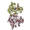
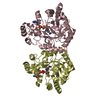
| Title | Crystal structure of apo form of Xylose reductase from Debaryomyces nepalensis | ||||||
 Components Components | Aldose reductase | ||||||
 Keywords Keywords | OXIDOREDUCTASE / carbohydrate metabolism / aldo-keto reductase / TIM barrel / NADPH dependent | ||||||
| Function / homology |  Function and homology information Function and homology informationD-xylose reductase [NAD(P)H] / D-xylose reductase (NADPH) activity / D-xylose catabolic process / nucleotide binding Similarity search - Function | ||||||
| Biological species |  Debaryomyces nepalensis (yeast) Debaryomyces nepalensis (yeast) | ||||||
| Method |  X-RAY DIFFRACTION / MOLECULAR REPLACEMENT / Resolution: 2 Å X-RAY DIFFRACTION / MOLECULAR REPLACEMENT / Resolution: 2 Å | ||||||
 Authors Authors | Manoj, N. | ||||||
 Citation Citation | Journal: FEBS J. / Year: 2018 Title: Crystal structure of yeast xylose reductase in complex with a novel NADP-DTT adduct provides insights into substrate recognition and catalysis. Authors: Paidimuddala, B. / Mohapatra, S.B. / Gummadi, S.N. / Manoj, N. #1: Journal: RSC Adv. / Year: 2017Title: A halotolerant aldose reductase from Debaryomyces nepalensis: gene isolation, overexpression and biochemical characterization Authors: Paidimuddala, B. / Aradhyam, G.K. / Gummadi, S.N. | ||||||
| History |
|
- Structure visualization
| Structure viewer | Molecule: MolmilJmol/JSmol |
|---|
- Downloads & links
-Download
| PDBx/mmCIF format | 5zci.cif.gz | 246.7 KB | Display | PDBx/mmCIF format |
|---|---|---|---|---|
| PDB format | pdb5zci.ent.gz | 197.5 KB | Display | PDB format |
| PDBx/mmJSON format | 5zci.json.gz | Tree view | PDBx/mmJSON format | |
| Others |  Other downloads Other downloads |
-Validation report
| Arichive directory | https://data.pdbj.org/pub/pdb/validation_reports/zc/5zciftp://data.pdbj.org/pub/pdb/validation_reports/zc/5zci | HTTPS FTP |
|---|
-Related structure data
| Related structure data |  5zcmC  1jezS S: Starting model for refinement C: citing same article ( |
|---|---|
| Similar structure data |

-Links
 PDBj
PDBj
- Assembly
| Deposited unit | 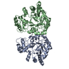
| ||||||||
|---|---|---|---|---|---|---|---|---|---|
| 1 |
| ||||||||
| Unit cell |
|
-Components
| #1: Protein | Mass: 38920.312 Da / Num. of mol.: 2 Source method: isolated from a genetically manipulated source Source: (gene. exp.) Debaryomyces nepalensis (yeast) / Gene: AR / Plasmid: pET28a / Production host:  #2: Water | ChemComp-HOH / |  Mass: 18.015 Da / Num. of mol.: 399 / Source method: isolated from a natural source / Formula: H2O Mass: 18.015 Da / Num. of mol.: 399 / Source method: isolated from a natural source / Formula: H2O |
|---|
-Experimental details
-Experiment
| Experiment | Method: X-RAY DIFFRACTION / Number of used crystals: 1 |
|---|
- Sample preparation
| Crystal | Density Matthews: 2.2 Å3/Da / Density % sol: 44.1 % |
|---|---|
| Crystal grow | Temperature: 293 K / Method: vapor diffusion, hanging drop / pH: 6.2 Details: 24% PEG 3000, 0.1 M sodium citrate (pH 6.2), 0.15 M ammonium acetate |
-Data collection
| Diffraction | Mean temperature: 100 K |
|---|---|
| Diffraction source | Source: ROTATING ANODE / Type: BRUKER AXS MICROSTAR / Wavelength: 1.5418 Å |
| Detector | Type: MAR scanner 345 mm plate / Detector: IMAGE PLATE / Date: Feb 3, 2016 |
| Radiation | Monochromator: double mirrors / Protocol: SINGLE WAVELENGTH / Monochromatic (M) / Laue (L): M / Scattering type: x-ray |
| Radiation wavelength | Wavelength: 1.5418 Å / Relative weight: 1 |
| Reflection | Resolution: 2→52.4 Å / Num. obs: 42060 / % possible obs: 89.2 % / Observed criterion σ(F): 0 / Observed criterion σ(I): 0 / Redundancy: 10.5 % / Biso Wilson estimate: 25.9 Å2 / CC1/2: 0.997 / Rmerge(I) obs: 0.157 / Rpim(I) all: 0.049 / Rrim(I) all: 0.165 / Net I/σ(I): 12.6 |
| Reflection shell | Resolution: 2→2.11 Å / Redundancy: 10.7 % / Rmerge(I) obs: 0.745 / Mean I/σ(I) obs: 3.1 / Num. unique obs: 5483 / CC1/2: 0.779 / Rpim(I) all: 0.232 / Rrim(I) all: 0.781 / % possible all: 81.1 |
- Processing
| Software |
| ||||||||||||||||||||||||
|---|---|---|---|---|---|---|---|---|---|---|---|---|---|---|---|---|---|---|---|---|---|---|---|---|---|
| Refinement | Method to determine structure: MOLECULAR REPLACEMENT Starting model: 1JEZ Resolution: 2→48.857 Å / SU ML: 0.22 / SU R Cruickshank DPI: 0.224 / Cross valid method: FREE R-VALUE / σ(F): 1.35 / Phase error: 20.74
| ||||||||||||||||||||||||
| Solvent computation | Shrinkage radii: 0.9 Å / VDW probe radii: 1.11 Å | ||||||||||||||||||||||||
| Displacement parameters | Biso mean: 27.2 Å2 | ||||||||||||||||||||||||
| Refine analyze | Luzzati coordinate error obs: 0.238 Å / Luzzati d res low obs: 48.9 Å | ||||||||||||||||||||||||
| Refinement step | Cycle: LAST / Resolution: 2→48.857 Å
| ||||||||||||||||||||||||
| Refine LS restraints |
| ||||||||||||||||||||||||
| LS refinement shell | Resolution: 2→2.047 Å
|
