 Movie
Movie Controller


+ Open data

- Basic information
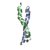
| Entry | Database: PDB / ID: 1jes | ||||||||||||||||||
|---|---|---|---|---|---|---|---|---|---|---|---|---|---|---|---|---|---|---|---|
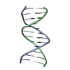
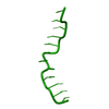
| Title | Crystal Structure of a Copper-Mediated Base Pair in DNA | ||||||||||||||||||
 Components Components | 5'-D(* Keywords KeywordsDNA / Cu / copper / Z-DNA / dodecamer / duplex / unnatural / designed | Function / homology | COPPER (II) ION / DNA / DNA (> 10) |  Function and homology information Function and homology informationMethod |  X-RAY DIFFRACTION / SYNCHROTRON / MAD / Resolution: 1.5 Å X-RAY DIFFRACTION / SYNCHROTRON / MAD / Resolution: 1.5 Å  Authors AuthorsAtwell, S. / Meggers, E. / Spraggon, G. / Schultz, P.G. |  CitationJournal: J.Am.Chem.Soc. / Year: 2001 CitationJournal: J.Am.Chem.Soc. / Year: 2001Title: Structure of a Copper-Mediated Base Pair in DNA Authors: Atwell, S. / Meggers, E. / Spraggon, G. / Schultz, P.G. #1: Journal: J.Am.Chem.Soc. / Year: 2000Title: A Novel Copper-Mediated DNA Base Pair Authors: Meggers, E. / Holland, P.L. / Tolman, W.B. / Romesberg, F.E. / Schultz, P.G. #2: Journal: Science / Year: 1981Title: Left-handed double helical DNA: variations in the backbone conformation Authors: Wang, A.G.-J. / Quigley, G.J. / Kolpak, F.J. / van Der Marel, G. / van Boom, J.H. / Rich, A. #3: Journal: Proc.Natl.Acad.Sci.USA / Year: 1981Title: Structure of a B-DNA dodecamer: conformation and dynamics Authors: Drew, H.R. / Wing, R.M. / Takano, T. / Broka, C. / Tanaka, S. / Itakura, K. / Dickerson, R.E. History |
|
- Structure visualization
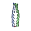
| Structure viewer | Molecule: MolmilJmol/JSmol |
|---|
- Downloads & links
-Download
| PDBx/mmCIF format | 1jes.cif.gz | 25.9 KB | Display | PDBx/mmCIF format |
|---|---|---|---|---|
| PDB format | pdb1jes.ent.gz | 17.1 KB | Display | PDB format |
| PDBx/mmJSON format | 1jes.json.gz | Tree view | PDBx/mmJSON format | |
| Others |  Other downloads Other downloads |
-Validation report
| Arichive directory | https://data.pdbj.org/pub/pdb/validation_reports/je/1jesftp://data.pdbj.org/pub/pdb/validation_reports/je/1jes | HTTPS FTP |
|---|
-Related structure data
| Related structure data | |
|---|---|
| Similar structure data |


-Links
 PDBj
PDBj


- Assembly
| Deposited unit | 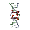
| ||||||||
|---|---|---|---|---|---|---|---|---|---|
| 1 |
| ||||||||
| Unit cell |
|
-Components
| #1: DNA chain | Mass: 3648.371 Da / Num. of mol.: 2 / Source method: obtained synthetically #2: Chemical |   Mass: 63.546 Da / Num. of mol.: 2 / Source method: obtained synthetically / Formula: Cu Mass: 63.546 Da / Num. of mol.: 2 / Source method: obtained synthetically / Formula: Cu#3: Water | ChemComp-HOH / |  Mass: 18.015 Da / Num. of mol.: 82 / Source method: isolated from a natural source / Formula: H2O Mass: 18.015 Da / Num. of mol.: 82 / Source method: isolated from a natural source / Formula: H2O |
|---|
-Experimental details
-Experiment
| Experiment | Method: X-RAY DIFFRACTION / Number of used crystals: 1 |
|---|
- Sample preparation
| Crystal | Density Matthews: 1.82 Å3/Da / Density % sol: 32.43 % | ||||||||||||||||||||||||
|---|---|---|---|---|---|---|---|---|---|---|---|---|---|---|---|---|---|---|---|---|---|---|---|---|---|
| Crystal grow | Temperature: 298 K / Method: vapor diffusion, hanging drop / pH: 7 Details: 10% MPD, 40 mM Na Cacodylate, 12 mM Spermine tetra-HCl, 80 mM NaCl, 20 mM MgCl2, pH 7.0, VAPOR DIFFUSION, HANGING DROP, temperature 298K | ||||||||||||||||||||||||
| Components of the solutions |
|
-Data collection
| Diffraction | Mean temperature: 140 K | ||||||||||||
|---|---|---|---|---|---|---|---|---|---|---|---|---|---|
| Diffraction source | Source: SYNCHROTRON / Site: ALS  / Beamline: 5.0.2 / Wavelength: 1.37991,1.37791,1.0 / Beamline: 5.0.2 / Wavelength: 1.37991,1.37791,1.0 | ||||||||||||
| Detector | Type: ADSC QUANTUM 4 / Detector: CCD / Date: Apr 6, 2001 / Details: Curved Si (111) | ||||||||||||
| Radiation | Protocol: MAD / Monochromatic (M) / Laue (L): M / Scattering type: x-ray | ||||||||||||
| Radiation wavelength |
| ||||||||||||
| Reflection | Resolution: 1.5→50 Å / Num. all: 47272 / Num. obs: 47190 / % possible obs: 98.2 % / Observed criterion σ(F): 0 / Observed criterion σ(I): 0 / Redundancy: 5.5 % / Biso Wilson estimate: 14.058 Å2 / Rsym value: 0.096 / Net I/σ(I): 46.7 | ||||||||||||
| Reflection shell | Resolution: 1.49→1.54 Å / Rmerge(I) obs: 0.686 / % possible all: 84.1 | ||||||||||||
| Reflection | *PLUS Num. obs: 8531 / Num. measured all: 47272 / Rmerge(I) obs: 0.096 | ||||||||||||
| Reflection shell | *PLUS % possible obs: 84.1 % / Rmerge(I) obs: 0.686 |
- Processing
| Software |
| |||||||||||||||||||||||||
|---|---|---|---|---|---|---|---|---|---|---|---|---|---|---|---|---|---|---|---|---|---|---|---|---|---|---|
| Refinement | Method to determine structure: MAD Starting model: initial map fit with mononucleotides Resolution: 1.5→50 Å / Cross valid method: free r / σ(F): 0 / σ(I): 0 Stereochemistry target values: G. Parkinson, J. Vojtechovsky, L. Clowney, A.T. Brunger, H.M. Berman, New Parameters for the Refinement of Nucleic Acid Containing Structures, Acta Cryst. D, 52, 57-64 (1996). Details: The fofc maps indicated an alternate conformation for the phosphate of B23, and so the structure was refined with two conformations for the phosphate of B23 (excepting the O5') and most of ...Details: The fofc maps indicated an alternate conformation for the phosphate of B23, and so the structure was refined with two conformations for the phosphate of B23 (excepting the O5') and most of residue B22 (excepting O5'), each with 0.5 occupancy.
| |||||||||||||||||||||||||
| Displacement parameters |
| |||||||||||||||||||||||||
| Refinement step | Cycle: LAST / Resolution: 1.5→50 Å
| |||||||||||||||||||||||||
| Refine LS restraints |
| |||||||||||||||||||||||||
| Software | *PLUS Name: CNS / Classification: refinement | |||||||||||||||||||||||||
| Refinement | *PLUS Highest resolution: 1.5 Å / Lowest resolution: 50 Å / Num. reflection obs: 8531 / σ(F): 0 / % reflection Rfree: 8 % / Rfactor obs: 0.169 | |||||||||||||||||||||||||
| Solvent computation | *PLUS | |||||||||||||||||||||||||
| Displacement parameters | *PLUS |
