 Movie
Movie Controller

[English] 日本語
 Yorodumi
- PDB-1je1: 5'-DEOXY-5'-METHYLTHIOADENOSINE PHOSPHORYLASE COMPLEX WITH GUANOS... -
+ Open data

- Basic information
| Entry | Database: PDB / ID: 1je1 | ||||||
|---|---|---|---|---|---|---|---|
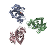
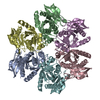
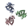
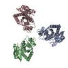
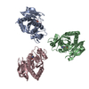
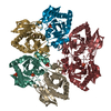
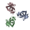
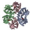
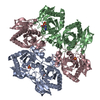
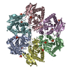
| Title | 5'-DEOXY-5'-METHYLTHIOADENOSINE PHOSPHORYLASE COMPLEX WITH GUANOSINE AND SULFATE | ||||||
 Components Components | 5'-METHYLTHIOADENOSINE PHOSPHORYLASE | ||||||
 Keywords Keywords | TRANSFERASE / alpha-beta protein | ||||||
| Function / homology |  Function and homology information Function and homology informationS-methyl-5'-thioadenosine phosphorylase / S-methyl-5-thioadenosine phosphorylase activity / nucleoside catabolic process / purine-nucleoside phosphorylase / cytosol Similarity search - Function | ||||||
| Biological species |   Sulfolobus solfataricus (archaea) Sulfolobus solfataricus (archaea) | ||||||
| Method |  X-RAY DIFFRACTION / SYNCHROTRON / MOLECULAR REPLACEMENT / Resolution: 1.8 Å X-RAY DIFFRACTION / SYNCHROTRON / MOLECULAR REPLACEMENT / Resolution: 1.8 Å | ||||||
 Authors Authors | Appleby, T.C. / Mathews, I.I. / Porcelli, M. / Cacciapuoti, G. / Ealick, S.E. | ||||||
 Citation Citation | Journal: J.Biol.Chem. / Year: 2001 Title: Three-dimensional structure of a hyperthermophilic 5'-deoxy-5'-methylthioadenosine phosphorylase from Sulfolobus solfataricus. Authors: Appleby, T.C. / Mathews, I.I. / Porcelli, M. / Cacciapuoti, G. / Ealick, S.E. #1: Journal: J.Biol.Chem. / Year: 1994Title: Purification and characterization of extremely thermophilic and thermostable 5'-methylthioadenosine phosphorylase from the archaeon Sulfolobus solfataricus. Purine nucleoside phosphorylase ...Title: Purification and characterization of extremely thermophilic and thermostable 5'-methylthioadenosine phosphorylase from the archaeon Sulfolobus solfataricus. Purine nucleoside phosphorylase activity and evidence for intersubunit disulfide bonds Authors: Cacciapuoti, G. / Porcelli, M. / Bertoldo, C. / De Rosa, M. / Zappia, V. #2: Journal: J.Biol.Chem. / Year: 1990Title: Three-Dimensional Structure of Human Erythrocytic Purine Nucleoside Phosphorylase At 3.2 Resolution Authors: Ealick, S.E. / Rule, S.A. / Carter, D.C. / Greenhough, T.J. / Babu, Y.S. #3: Journal: Structure / Year: 1999Title: The structure of human 5'-deoxy-5'-methylthioadenosine phosphorylase at 1.7 resolution provides insights into substrate binding and catalysis Authors: Appleby, T.C. / Erion, M.D. / Ealick, S.E. | ||||||
| History |
|
- Structure visualization
| Structure viewer | Molecule: MolmilJmol/JSmol |
|---|
- Downloads & links
-Download
| PDBx/mmCIF format | 1je1.cif.gz | 278.7 KB | Display | PDBx/mmCIF format |
|---|---|---|---|---|
| PDB format | pdb1je1.ent.gz | 226.9 KB | Display | PDB format |
| PDBx/mmJSON format | 1je1.json.gz | Tree view | PDBx/mmJSON format | |
| Others |  Other downloads Other downloads |
-Validation report
| Arichive directory | https://data.pdbj.org/pub/pdb/validation_reports/je/1je1ftp://data.pdbj.org/pub/pdb/validation_reports/je/1je1 | HTTPS FTP |
|---|
-Related structure data
| Related structure data |  1jdsC  1jdtC  1jduC  1jdvC  1jdzC  1je0C  1jp7C  1jpvC C: citing same article ( |
|---|---|
| Similar structure data |
-Links
 PDBj
PDBj- Assembly
| Deposited unit | 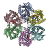
| ||||||||||
|---|---|---|---|---|---|---|---|---|---|---|---|
| 1 |
| ||||||||||
| Unit cell |
|
-Components
| #1: Protein | Mass: 25763.457 Da / Num. of mol.: 6 Source method: isolated from a genetically manipulated source Source: (gene. exp.) Sulfolobus solfataricus (archaea) / Production host:  References: UniProt: P50389, S-methyl-5'-thioadenosine phosphorylase #2: Chemical | ChemComp-SO4 /   Mass: 96.063 Da / Num. of mol.: 6 / Source method: obtained synthetically / Formula: SO4 Mass: 96.063 Da / Num. of mol.: 6 / Source method: obtained synthetically / Formula: SO4#3: Chemical | ChemComp-GMP /   Mass: 283.241 Da / Num. of mol.: 6 / Source method: obtained synthetically / Formula: C10H13N5O5 Mass: 283.241 Da / Num. of mol.: 6 / Source method: obtained synthetically / Formula: C10H13N5O5#4: Water | ChemComp-HOH / |  Mass: 18.015 Da / Num. of mol.: 417 / Source method: isolated from a natural source / Formula: H2O Mass: 18.015 Da / Num. of mol.: 417 / Source method: isolated from a natural source / Formula: H2OHas protein modification | Y | |
|---|
-Experimental details
-Experiment
| Experiment | Method: X-RAY DIFFRACTION / Number of used crystals: 1 |
|---|
- Sample preparation
| Crystal | Density Matthews: 2.54 Å3/Da / Density % sol: 51.62 % | ||||||||||||||||||||||||||||||||||||||||||
|---|---|---|---|---|---|---|---|---|---|---|---|---|---|---|---|---|---|---|---|---|---|---|---|---|---|---|---|---|---|---|---|---|---|---|---|---|---|---|---|---|---|---|---|
| Crystal grow | Temperature: 291 K / Method: vapor diffusion, hanging drop / pH: 7.4 Details: dioxane, MPD, MgCl2, NaCl, Tris.HCl, pH 7.4, VAPOR DIFFUSION, HANGING DROP at 291K | ||||||||||||||||||||||||||||||||||||||||||
| Crystal grow | *PLUS Details: used microseeding | ||||||||||||||||||||||||||||||||||||||||||
| Components of the solutions | *PLUS
|
-Data collection
| Diffraction | Mean temperature: 100 K |
|---|---|
| Diffraction source | Source: SYNCHROTRON / Site: CHESS  / Beamline: A1 / Wavelength: 0.9023 Å / Beamline: A1 / Wavelength: 0.9023 Å |
| Detector | Type: ADSC QUANTUM 4 / Detector: CCD |
| Radiation | Monochromator: Si(111) / Protocol: SINGLE WAVELENGTH / Monochromatic (M) / Laue (L): M / Scattering type: x-ray |
| Radiation wavelength | Wavelength: 0.9023 Å / Relative weight: 1 |
| Reflection | Resolution: 1.8→30 Å / Num. all: 139268 / Num. obs: 139268 / % possible obs: 82 % / Observed criterion σ(I): 1 / Redundancy: 3.9 % / Rmerge(I) obs: 0.057 / Net I/σ(I): 7.4 |
| Reflection shell | Highest resolution: 1.8 Å / Rmerge(I) obs: 0.316 / Mean I/σ(I) obs: 2.3 / % possible all: 41.7 |
| Reflection | *PLUS Num. measured all: 543683 |
| Reflection shell | *PLUS % possible obs: 41.7 % |
- Processing
| Software |
| |||||||||||||||||||||||||
|---|---|---|---|---|---|---|---|---|---|---|---|---|---|---|---|---|---|---|---|---|---|---|---|---|---|---|
| Refinement | Method to determine structure: MOLECULAR REPLACEMENT Starting model: native complex Resolution: 1.8→20 Å / Cross valid method: THROUGHOUT / σ(F): 2 / σ(I): 1 / Stereochemistry target values: Engh & Huber
| |||||||||||||||||||||||||
| Displacement parameters | Biso mean: 28.9 Å2 | |||||||||||||||||||||||||
| Refinement step | Cycle: LAST / Resolution: 1.8→20 Å
| |||||||||||||||||||||||||
| Refine LS restraints |
| |||||||||||||||||||||||||
| Software | *PLUS Name: CNS / Version: 0.9 / Classification: refinement | |||||||||||||||||||||||||
| Refinement | *PLUS Highest resolution: 1.8 Å / Lowest resolution: 20 Å / σ(F): 2 / % reflection Rfree: 5.1 % / Rfactor obs: 0.217 | |||||||||||||||||||||||||
| Solvent computation | *PLUS | |||||||||||||||||||||||||
| Displacement parameters | *PLUS Biso mean: 28.9 Å2 |
