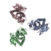
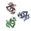
 Movie
Movie Controller


+ Open data

- Basic information
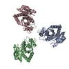
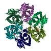
| Entry | Database: PDB / ID: 1ov6 | ||||||
|---|---|---|---|---|---|---|---|
| Title | M64V PNP + ALLO | ||||||
 Components Components | Purine nucleoside phosphorylase | ||||||
 Keywords Keywords | TRANSFERASE / M64V / mutant PNP / ALLO | ||||||
| Function / homology |  Function and homology information Function and homology informationpurine nucleoside interconversion / guanosine phosphorylase activity / purine-nucleoside phosphorylase / purine-nucleoside phosphorylase activity / purine nucleoside catabolic process / DNA damage response / membrane / identical protein binding / cytosol Similarity search - Function | ||||||
| Biological species |  | ||||||
| Method |  X-RAY DIFFRACTION / SYNCHROTRON / MOLECULAR REPLACEMENT / Resolution: 2.4 Å X-RAY DIFFRACTION / SYNCHROTRON / MOLECULAR REPLACEMENT / Resolution: 2.4 Å | ||||||
 Authors Authors | Ealick, S.E. / Bennett, E.M. / Anand, R. / Secrist, J.A. / Parker, P.W. / Hassan, A.E. / Allan, P.W. / McPherson, D.T. / Sorscher, E.J. | ||||||
 Citation Citation | Journal: Chem.Biol. / Year: 2003 Title: Designer gene therapy using an Escherichia coli purine nucleoside phosphorylase/prodrug system. Authors: Bennett, E.M. / Anand, R. / Allan, P.W. / Hassan, A.E. / Hong, J.S. / Levasseur, D.N. / McPherson, D.T. / Parker, W.B. / Secrist, J.A. / Sorscher, E.J. / Townes, T.M. / Waud, W.R. / Ealick, S.E. | ||||||
| History |
|
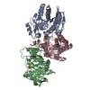
- Structure visualization
| Structure viewer | Molecule: MolmilJmol/JSmol |
|---|
- Downloads & links
-Download
| PDBx/mmCIF format | 1ov6.cif.gz | 147 KB | Display | PDBx/mmCIF format |
|---|---|---|---|---|
| PDB format | pdb1ov6.ent.gz | 117.8 KB | Display | PDB format |
| PDBx/mmJSON format | 1ov6.json.gz | Tree view | PDBx/mmJSON format | |
| Others |  Other downloads Other downloads |
-Validation report
| Arichive directory | https://data.pdbj.org/pub/pdb/validation_reports/ov/1ov6ftp://data.pdbj.org/pub/pdb/validation_reports/ov/1ov6 | HTTPS FTP |
|---|
-Related structure data


-Links
 PDBj
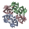
PDBj- Assembly
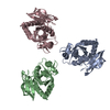
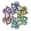
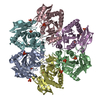
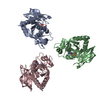
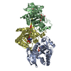
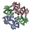
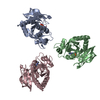
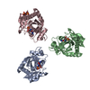
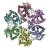
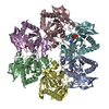
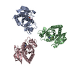
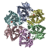
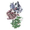
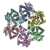
| Deposited unit | 
| ||||||||
|---|---|---|---|---|---|---|---|---|---|
| 1 | 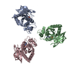
| ||||||||
| Unit cell |
|
-Components
| #1: Protein | Mass: 25818.684 Da / Num. of mol.: 3 / Fragment: Purine Nuleoside Phosphorylase / Mutation: M64V Source method: isolated from a genetically manipulated source Source: (gene. exp.) References: UniProt: P0ABP8, purine-nucleoside phosphorylase #2: Chemical |   Mass: 94.971 Da / Num. of mol.: 3 / Source method: obtained synthetically / Formula: PO4 Mass: 94.971 Da / Num. of mol.: 3 / Source method: obtained synthetically / Formula: PO4#3: Chemical | 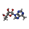  Type: RNA linking / Mass: 280.280 Da / Num. of mol.: 3 / Source method: obtained synthetically / Formula: C12H16N4O4 Type: RNA linking / Mass: 280.280 Da / Num. of mol.: 3 / Source method: obtained synthetically / Formula: C12H16N4O4#4: Water | ChemComp-HOH / |  Mass: 18.015 Da / Num. of mol.: 185 / Source method: isolated from a natural source / Formula: H2O Mass: 18.015 Da / Num. of mol.: 185 / Source method: isolated from a natural source / Formula: H2O |
|---|
-Experimental details
-Experiment
| Experiment | Method: X-RAY DIFFRACTION / Number of used crystals: 1 |
|---|
- Sample preparation
| Crystal | Density Matthews: 3.26 Å3/Da / Density % sol: 62.3 % | |||||||||||||||||||||||||||||||||||
|---|---|---|---|---|---|---|---|---|---|---|---|---|---|---|---|---|---|---|---|---|---|---|---|---|---|---|---|---|---|---|---|---|---|---|---|---|
| Crystal grow | Temperature: 295 K / Method: vapor diffusion, hanging drop / pH: 5.4 Details: 50mM sodium citrate, 30% ammonium sulfate, pH 5.4, VAPOR DIFFUSION, HANGING DROP, temperature 295K | |||||||||||||||||||||||||||||||||||
| Crystal grow | *PLUS pH: 8 / Method: vapor diffusion, hanging drop | |||||||||||||||||||||||||||||||||||
| Components of the solutions | *PLUS
|
-Data collection
| Diffraction | Mean temperature: 170 K |
|---|---|
| Diffraction source | Source: SYNCHROTRON / Site: CHESS  / Beamline: F1 / Wavelength: 0.948 Å / Beamline: F1 / Wavelength: 0.948 Å |
| Detector | Type: ADSC QUANTUM 4 / Detector: CCD / Date: Dec 25, 2001 |
| Radiation | Protocol: SINGLE WAVELENGTH / Monochromatic (M) / Laue (L): M / Scattering type: x-ray |
| Radiation wavelength | Wavelength: 0.948 Å / Relative weight: 1 |
| Reflection | Resolution: 2.4→25 Å / Num. all: 41133 / Num. obs: 41133 / % possible obs: 100 % / Observed criterion σ(F): 0 / Observed criterion σ(I): 0 / Redundancy: 10 % / Biso Wilson estimate: 26 Å2 / Rmerge(I) obs: 0.11 / Rsym value: 0.11 / Net I/σ(I): 4.5 |
| Reflection shell | Resolution: 2.4→2.55 Å / Redundancy: 10 % / Rmerge(I) obs: 0.332 / Mean I/σ(I) obs: 2.1 / Rsym value: 0.332 / % possible all: 100 |
| Reflection | *PLUS Redundancy: 10 % / Rmerge(I) obs: 0.11 |
| Reflection shell | *PLUS % possible obs: 100 % |
- Processing
| Software |
| |||||||||||||||||||||||||
|---|---|---|---|---|---|---|---|---|---|---|---|---|---|---|---|---|---|---|---|---|---|---|---|---|---|---|
| Refinement | Method to determine structure: MOLECULAR REPLACEMENT / Resolution: 2.4→24.82 Å / Rfactor Rfree error: 0.004 / Isotropic thermal model: RESTRAINED / Cross valid method: THROUGHOUT / σ(F): 0 / σ(I): 0 / Stereochemistry target values: Engh & Huber
| |||||||||||||||||||||||||
| Solvent computation | Solvent model: FLAT MODEL / Bsol: 50.9449 Å2 / ksol: 0.447286 e/Å3 | |||||||||||||||||||||||||
| Displacement parameters | Biso mean: 25.2 Å2
| |||||||||||||||||||||||||
| Refine analyze |
| |||||||||||||||||||||||||
| Refinement step | Cycle: LAST / Resolution: 2.4→24.82 Å
| |||||||||||||||||||||||||
| Refine LS restraints |
| |||||||||||||||||||||||||
| LS refinement shell | Resolution: 2.4→2.55 Å / Rfactor Rfree error: 0.01 / Total num. of bins used: 6
| |||||||||||||||||||||||||
| Xplor file |
| |||||||||||||||||||||||||
| Refinement | *PLUS Highest resolution: 2.4 Å / Lowest resolution: 25 Å / Rfactor Rwork: 0.23 | |||||||||||||||||||||||||
| Solvent computation | *PLUS | |||||||||||||||||||||||||
| Displacement parameters | *PLUS | |||||||||||||||||||||||||
| Refine LS restraints | *PLUS
|
