 Movie
Movie Controller

[English] 日本語
 Yorodumi
- PDB-4rj2: Crystal structure of E.coli purine nucleoside phosphorylase at 0.... -
+ Open data

- Basic information
| Entry | Database: PDB / ID: 4rj2 | ||||||
|---|---|---|---|---|---|---|---|
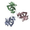
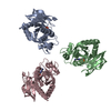
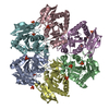
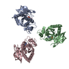
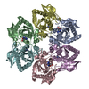
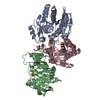
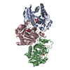
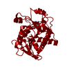
| Title | Crystal structure of E.coli purine nucleoside phosphorylase at 0.99 A resolution | ||||||
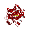
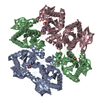
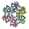
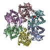
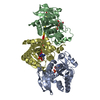
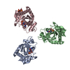
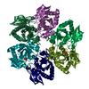
 Components Components | Purine nucleoside phosphorylase DeoD-type | ||||||
 Keywords Keywords | TRANSFERASE / phosphorylase | ||||||
| Function / homology | Nucleoside phosphorylase domain / Rossmann fold / 3-Layer(aba) Sandwich / Alpha Beta / :  Function and homology information Function and homology information | ||||||
| Biological species |  | ||||||
| Method |  X-RAY DIFFRACTION / SYNCHROTRON / MOLECULAR REPLACEMENT / Resolution: 0.99 Å X-RAY DIFFRACTION / SYNCHROTRON / MOLECULAR REPLACEMENT / Resolution: 0.99 Å | ||||||
 Authors Authors | Timofeev, V.I. / Abramchik, Y.A. / Esipov, R.S. / Kuranova, I.P. | ||||||
 Citation Citation | Journal: To be Published Title: Crystal structure of E.coli purine nucleoside phosphorylase at 0.99 A resolution Authors: Timofeev, V.I. / Abramchik, Y.A. / Esipov, R.S. / Kuranova, I.P. | ||||||
| History |
|
- Structure visualization
| Structure viewer | Molecule: MolmilJmol/JSmol |
|---|
- Downloads & links
-Download
| PDBx/mmCIF format | 4rj2.cif.gz | 575.3 KB | Display | PDBx/mmCIF format |
|---|---|---|---|---|
| PDB format | pdb4rj2.ent.gz | 478.8 KB | Display | PDB format |
| PDBx/mmJSON format | 4rj2.json.gz | Tree view | PDBx/mmJSON format | |
| Others |  Other downloads Other downloads |
-Validation report
| Arichive directory | https://data.pdbj.org/pub/pdb/validation_reports/rj/4rj2ftp://data.pdbj.org/pub/pdb/validation_reports/rj/4rj2 | HTTPS FTP |
|---|
-Related structure data
| Similar structure data |
|---|

-Links
 PDBj
PDBj- Assembly
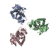
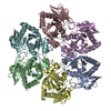
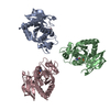
| Deposited unit | 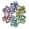
| ||||||||
|---|---|---|---|---|---|---|---|---|---|
| 1 |
| ||||||||
| Unit cell |
|
-Components
| #1: Protein | Mass: 25721.633 Da / Num. of mol.: 6 Source method: isolated from a genetically manipulated source Source: (gene. exp.) References: UniProt: H0QGF5, purine-nucleoside phosphorylase #2: Chemical | ChemComp-GOL /   Mass: 92.094 Da / Num. of mol.: 6 / Source method: obtained synthetically / Formula: C3H8O3 Mass: 92.094 Da / Num. of mol.: 6 / Source method: obtained synthetically / Formula: C3H8O3#3: Water | ChemComp-HOH / |  Mass: 18.015 Da / Num. of mol.: 949 / Source method: isolated from a natural source / Formula: H2O Mass: 18.015 Da / Num. of mol.: 949 / Source method: isolated from a natural source / Formula: H2O |
|---|
-Experimental details
-Experiment
| Experiment | Method: X-RAY DIFFRACTION / Number of used crystals: 1 |
|---|
- Sample preparation
| Crystal | Density Matthews: 2.18 Å3/Da / Density % sol: 43.54 % |
|---|---|
| Crystal grow | Method: counter-diffusion / Details: COUNTER DIFFUSION |
-Data collection
| Diffraction | Mean temperature: 100 K |
|---|---|
| Diffraction source | Source: SYNCHROTRON / Site: SPring-8  / Beamline: BL41XU / Wavelength: 0.8 Å / Beamline: BL41XU / Wavelength: 0.8 Å |
| Detector | Type: MARMOSAIC 225 mm CCD / Detector: CCD / Date: May 23, 2012 |
| Radiation | Protocol: SINGLE WAVELENGTH / Monochromatic (M) / Laue (L): M / Scattering type: x-ray |
| Radiation wavelength | Wavelength: 0.8 Å / Relative weight: 1 |
| Reflection | Resolution: 0.99→20 Å / Num. obs: 718472 / % possible obs: 98.41 % / Redundancy: 3.71 % / Rmerge(I) obs: 0.1 |
| Reflection shell | Resolution: 0.99→1.04 Å / Redundancy: 3.6 % / Rmerge(I) obs: 0.67 / Mean I/σ(I) obs: 2.11 / Num. unique all: 104431 / % possible all: 98.05 |
- Processing
| Software |
| ||||||||||||||||||||||||||||||||||||||||||||||||||||||||||||||||||||||||||||||||||||||||||||||||||||||||||||||||||||||||||||||||||||||||||||||||||||||||||||||||||||||||||||||||||||||
|---|---|---|---|---|---|---|---|---|---|---|---|---|---|---|---|---|---|---|---|---|---|---|---|---|---|---|---|---|---|---|---|---|---|---|---|---|---|---|---|---|---|---|---|---|---|---|---|---|---|---|---|---|---|---|---|---|---|---|---|---|---|---|---|---|---|---|---|---|---|---|---|---|---|---|---|---|---|---|---|---|---|---|---|---|---|---|---|---|---|---|---|---|---|---|---|---|---|---|---|---|---|---|---|---|---|---|---|---|---|---|---|---|---|---|---|---|---|---|---|---|---|---|---|---|---|---|---|---|---|---|---|---|---|---|---|---|---|---|---|---|---|---|---|---|---|---|---|---|---|---|---|---|---|---|---|---|---|---|---|---|---|---|---|---|---|---|---|---|---|---|---|---|---|---|---|---|---|---|---|---|---|---|---|
| Refinement | Method to determine structure: MOLECULAR REPLACEMENT / Resolution: 0.99→16.28 Å / Cor.coef. Fo:Fc: 0.967 / Cor.coef. Fo:Fc free: 0.964 / Cross valid method: THROUGHOUT / ESU R: 0.026 / ESU R Free: 0.026 / Stereochemistry target values: MAXIMUM LIKELIHOOD / Details: HYDROGENS HAVE BEEN ADDED IN THE RIDING POSITIONS
| ||||||||||||||||||||||||||||||||||||||||||||||||||||||||||||||||||||||||||||||||||||||||||||||||||||||||||||||||||||||||||||||||||||||||||||||||||||||||||||||||||||||||||||||||||||||
| Solvent computation | Ion probe radii: 0.8 Å / Shrinkage radii: 0.8 Å / VDW probe radii: 1.2 Å / Solvent model: MASK | ||||||||||||||||||||||||||||||||||||||||||||||||||||||||||||||||||||||||||||||||||||||||||||||||||||||||||||||||||||||||||||||||||||||||||||||||||||||||||||||||||||||||||||||||||||||
| Displacement parameters | Biso mean: 11.135 Å2
| ||||||||||||||||||||||||||||||||||||||||||||||||||||||||||||||||||||||||||||||||||||||||||||||||||||||||||||||||||||||||||||||||||||||||||||||||||||||||||||||||||||||||||||||||||||||
| Refinement step | Cycle: LAST / Resolution: 0.99→16.28 Å
| ||||||||||||||||||||||||||||||||||||||||||||||||||||||||||||||||||||||||||||||||||||||||||||||||||||||||||||||||||||||||||||||||||||||||||||||||||||||||||||||||||||||||||||||||||||||
| Refine LS restraints |
|