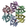
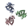
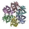
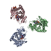
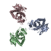
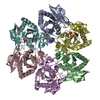
| 登録構造単位 | A: Purine nucleoside phosphorylase deoD-type
B: Purine nucleoside phosphorylase deoD-type
C: Purine nucleoside phosphorylase deoD-type
D: Purine nucleoside phosphorylase deoD-type
E: Purine nucleoside phosphorylase deoD-type
F: Purine nucleoside phosphorylase deoD-type
G: Purine nucleoside phosphorylase deoD-type
H: Purine nucleoside phosphorylase deoD-type
I: Purine nucleoside phosphorylase deoD-type
J: Purine nucleoside phosphorylase deoD-type
K: Purine nucleoside phosphorylase deoD-type
L: Purine nucleoside phosphorylase deoD-type
M: Purine nucleoside phosphorylase deoD-type
N: Purine nucleoside phosphorylase deoD-type
O: Purine nucleoside phosphorylase deoD-type
P: Purine nucleoside phosphorylase deoD-type
Q: Purine nucleoside phosphorylase deoD-type
R: Purine nucleoside phosphorylase deoD-type
ヘテロ分子
| 分子量 (理論値) | 分子数 |
|---|
| 合計 (水以外) | 465,174 | 41 |
|---|
| ポリマ- | 462,989 | 18 |
|---|
| 非ポリマー | 2,184 | 23 |
|---|
| 水 | 20,771 | 1153 |
|---|
|
|---|
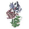
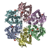
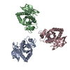
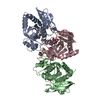
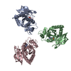
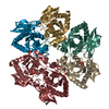
| 1 | A: Purine nucleoside phosphorylase deoD-type
B: Purine nucleoside phosphorylase deoD-type
C: Purine nucleoside phosphorylase deoD-type
D: Purine nucleoside phosphorylase deoD-type
E: Purine nucleoside phosphorylase deoD-type
F: Purine nucleoside phosphorylase deoD-type
ヘテロ分子
| 分子量 (理論値) | 分子数 |
|---|
| 合計 (水以外) | 154,995 | 13 |
|---|
| ポリマ- | 154,330 | 6 |
|---|
| 非ポリマー | 665 | 7 |
|---|
| 水 | 108 | 6 |
|---|
| タイプ | 名称 | 対称操作 | 数 |
|---|
| identity operation | 1_555 | x,y,z | 1 |
| Buried area | 20950 Å2 |
|---|
| ΔGint | -135 kcal/mol |
|---|
| Surface area | 44680 Å2 |
|---|
| 手法 | PISA |
|---|
|
|---|
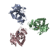
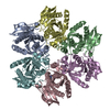
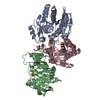
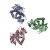
| 2 | G: Purine nucleoside phosphorylase deoD-type
H: Purine nucleoside phosphorylase deoD-type
I: Purine nucleoside phosphorylase deoD-type
J: Purine nucleoside phosphorylase deoD-type
K: Purine nucleoside phosphorylase deoD-type
L: Purine nucleoside phosphorylase deoD-type
ヘテロ分子
| 分子量 (理論値) | 分子数 |
|---|
| 合計 (水以外) | 155,185 | 15 |
|---|
| ポリマ- | 154,330 | 6 |
|---|
| 非ポリマー | 855 | 9 |
|---|
| 水 | 108 | 6 |
|---|
| タイプ | 名称 | 対称操作 | 数 |
|---|
| identity operation | 1_555 | x,y,z | 1 |
| Buried area | 21030 Å2 |
|---|
| ΔGint | -132 kcal/mol |
|---|
| Surface area | 44810 Å2 |
|---|
| 手法 | PISA |
|---|
|
|---|
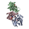
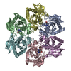
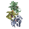
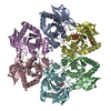
| 3 | M: Purine nucleoside phosphorylase deoD-type
N: Purine nucleoside phosphorylase deoD-type
O: Purine nucleoside phosphorylase deoD-type
P: Purine nucleoside phosphorylase deoD-type
Q: Purine nucleoside phosphorylase deoD-type
R: Purine nucleoside phosphorylase deoD-type
ヘテロ分子
| 分子量 (理論値) | 分子数 |
|---|
| 合計 (水以外) | 154,995 | 13 |
|---|
| ポリマ- | 154,330 | 6 |
|---|
| 非ポリマー | 665 | 7 |
|---|
| 水 | 108 | 6 |
|---|
| タイプ | 名称 | 対称操作 | 数 |
|---|
| identity operation | 1_555 | x,y,z | 1 |
| Buried area | 20970 Å2 |
|---|
| ΔGint | -132 kcal/mol |
|---|
| Surface area | 44760 Å2 |
|---|
| 手法 | PISA |
|---|
|
|---|
| 単位格子 | | Length a, b, c (Å) | 94.447, 165.923, 135.376 |
|---|
| Angle α, β, γ (deg.) | 90.000, 99.690, 90.000 |
|---|
| Int Tables number | 4 |
|---|
| Space group name H-M | P1211 |
|---|
|
|---|
 ムービー
ムービー コントローラー


 データを開く
 基本情報
基本情報 要素
要素 キーワード
キーワード 機能・相同性情報
 X線回折 /
  データ登録者
 引用
引用 構造の表示
構造の表示 ダウンロードとリンク
 その他のダウンロード


 PDBj
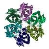
PDBj 集合体
集合体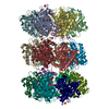

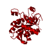
 分子量: 94.971 Da / 分子数: 23 / 由来タイプ: 合成 / 式: PO4
 分子量: 18.015 Da / 分子数: 1153 / 由来タイプ: 天然 / 式: H2O
 試料調製
試料調製 / ビームライン: BM14 / 波長: 0.9537 Å
 解析
解析