 ムービー
ムービー コントローラー


+ データを開く

- 基本情報
基本情報
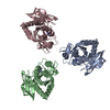
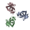
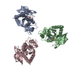
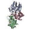
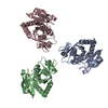
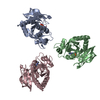
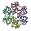
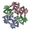
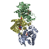
| 登録情報 | データベース: PDB / ID: 6xz2 | ||||||
|---|---|---|---|---|---|---|---|
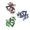
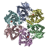
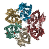
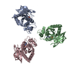
| タイトル | Crystal structure of E. Coli purine nucleoside phosphorylase mutant Y160W with SO4 and Formycin A | ||||||
 要素 要素 | Purine nucleoside phosphorylase DeoD-type | ||||||
 キーワード キーワード | TRANSFERASE / trimer of dimers / single tryptophan mutant / protein-inhibitor complex | ||||||
| 機能・相同性 |  機能・相同性情報 機能・相同性情報purine nucleoside interconversion / guanosine phosphorylase activity / purine-nucleoside phosphorylase / purine-nucleoside phosphorylase activity / purine nucleoside catabolic process / DNA damage response / membrane / identical protein binding / cytosol 類似検索 - 分子機能 | ||||||
| 生物種 |  | ||||||
| 手法 |  X線回折 / シンクロトロン / 分子置換 / 解像度: 1.65000304482 Å X線回折 / シンクロトロン / 分子置換 / 解像度: 1.65000304482 Å | ||||||
 データ登録者 データ登録者 | Narczyk, M. / Bzowska, A. | ||||||
 引用 引用 | ジャーナル: Sci Rep / 年: 2021 タイトル: Single tryptophan Y160W mutant of homooligomeric E. coli purine nucleoside phosphorylase implies that dimers forming the hexamer are functionally not equivalent. 著者: Narczyk, M. / Mioduszewski, L. / Oksiejuk, A. / Winiewska-Szajewska, M. / Wielgus-Kutrowska, B. / Gojdz, A. / Ciesla, J. / Bzowska, A. | ||||||
| 履歴 |
|
- 構造の表示
構造の表示
| 構造ビューア | 分子: MolmilJmol/JSmol |
|---|
- ダウンロードとリンク
-ダウンロード
| PDBx/mmCIF形式 | 6xz2.cif.gz | 185.5 KB | 表示 | PDBx/mmCIF形式 |
|---|---|---|---|---|
| PDB形式 | pdb6xz2.ent.gz | 118.9 KB | 表示 | PDB形式 |
| PDBx/mmJSON形式 | 6xz2.json.gz | ツリー表示 | PDBx/mmJSON形式 | |
| その他 |  その他のダウンロード その他のダウンロード |
-検証レポート
| アーカイブディレクトリ | https://data.pdbj.org/pub/pdb/validation_reports/xz/6xz2ftp://data.pdbj.org/pub/pdb/validation_reports/xz/6xz2 | HTTPS FTP |
|---|
-関連構造データ
| 関連構造データ |  1ecpS S: 精密化の開始モデル |
|---|---|
| 類似構造データ |
-リンク
 PDBj
PDBj- 集合体
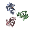
集合体
| 登録構造単位 | 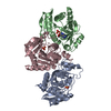
| |||||||||||||||
|---|---|---|---|---|---|---|---|---|---|---|---|---|---|---|---|---|
| 1 | 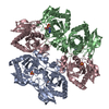
| |||||||||||||||
| 2 |
| |||||||||||||||
| 単位格子 |
| |||||||||||||||
| Components on special symmetry positions |
|
-要素
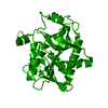
| #1: タンパク質 | 分子量: 25744.668 Da / 分子数: 3 / 変異: Y160W / 由来タイプ: 組換発現 由来: (組換発現) 株: K12 / 遺伝子: deoD, pup, b4384, JW4347 / 発現宿主: #2: 化合物 |   分子量: 96.063 Da / 分子数: 3 / 由来タイプ: 合成 / 式: SO4 分子量: 96.063 Da / 分子数: 3 / 由来タイプ: 合成 / 式: SO4#3: 化合物 | ChemComp-FMC / ( | 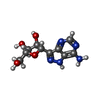  分子量: 267.241 Da / 分子数: 1 / 由来タイプ: 合成 / 式: C10H13N5O4 分子量: 267.241 Da / 分子数: 1 / 由来タイプ: 合成 / 式: C10H13N5O4#4: 水 | ChemComp-HOH / |  分子量: 18.015 Da / 分子数: 304 / 由来タイプ: 天然 / 式: H2O 分子量: 18.015 Da / 分子数: 304 / 由来タイプ: 天然 / 式: H2O研究の焦点であるリガンドがあるか | N | |
|---|
-実験情報
-実験
| 実験 | 手法: X線回折 / 使用した結晶の数: 1 |
|---|
- 試料調製
試料調製
| 結晶 | マシュー密度: 3.25 Å3/Da / 溶媒含有率: 62.18 % |
|---|---|
| 結晶化 | 温度: 291 K / 手法: 蒸気拡散法, ハンギングドロップ法 / pH: 5.2 詳細: 50 mM citrate buffer pH 5.2, 14 % ammonium sulphate (w/v) |
-データ収集
| 回折 | 平均測定温度: 100 K / Serial crystal experiment: N |
|---|---|
| 放射光源 | 由来: シンクロトロン / サイト: PETRA III, EMBL c/o DESY  / ビームライン: P14 (MX2) / 波長: 1.0064 Å / ビームライン: P14 (MX2) / 波長: 1.0064 Å |
| 検出器 | タイプ: DECTRIS EIGER X 16M / 検出器: PIXEL / 日付: 2019年11月28日 |
| 放射 | プロトコル: SINGLE WAVELENGTH / 単色(M)・ラウエ(L): M / 散乱光タイプ: x-ray |
| 放射波長 | 波長: 1.0064 Å / 相対比: 1 |
| 反射 | 解像度: 1.65→104.3 Å / Num. obs: 123021 / % possible obs: 99.42 % / 冗長度: 99.8 % / Biso Wilson estimate: 22.4977063367 Å2 / CC1/2: 0.998 / Rmerge(I) obs: 0.175 / Rpim(I) all: 0.028 / Rrim(I) all: 0.177 / Net I/σ(I): 14.7 |
| 反射 シェル | 解像度: 1.65→1.68 Å / Rmerge(I) obs: 1.483 / Num. unique obs: 5869 / CC1/2: 0.821 / Rpim(I) all: 0.26 / Rrim(I) all: 1.507 |
- 解析
解析
| ソフトウェア |
| |||||||||||||||||||||||||||||||||||||||||||||||||||||||||||||||||||||||||||||||||||||||||||||||||||||||||||||||||||||||||||||||||||||||||||||||||||||||||||||||||||||||||||||||||||||||||||||||||||||||||||||||||||||||||
|---|---|---|---|---|---|---|---|---|---|---|---|---|---|---|---|---|---|---|---|---|---|---|---|---|---|---|---|---|---|---|---|---|---|---|---|---|---|---|---|---|---|---|---|---|---|---|---|---|---|---|---|---|---|---|---|---|---|---|---|---|---|---|---|---|---|---|---|---|---|---|---|---|---|---|---|---|---|---|---|---|---|---|---|---|---|---|---|---|---|---|---|---|---|---|---|---|---|---|---|---|---|---|---|---|---|---|---|---|---|---|---|---|---|---|---|---|---|---|---|---|---|---|---|---|---|---|---|---|---|---|---|---|---|---|---|---|---|---|---|---|---|---|---|---|---|---|---|---|---|---|---|---|---|---|---|---|---|---|---|---|---|---|---|---|---|---|---|---|---|---|---|---|---|---|---|---|---|---|---|---|---|---|---|---|---|---|---|---|---|---|---|---|---|---|---|---|---|---|---|---|---|---|---|---|---|---|---|---|---|---|---|---|---|---|---|---|---|---|
| 精密化 | 構造決定の手法: 分子置換 開始モデル: 1ecp 解像度: 1.65000304482→78.7072722459 Å / SU ML: 0.157720237261 / 交差検証法: FREE R-VALUE / σ(F): 1.35096088804 / 位相誤差: 18.312385071
| |||||||||||||||||||||||||||||||||||||||||||||||||||||||||||||||||||||||||||||||||||||||||||||||||||||||||||||||||||||||||||||||||||||||||||||||||||||||||||||||||||||||||||||||||||||||||||||||||||||||||||||||||||||||||
| 溶媒の処理 | 減衰半径: 0.9 Å / VDWプローブ半径: 1.11 Å | |||||||||||||||||||||||||||||||||||||||||||||||||||||||||||||||||||||||||||||||||||||||||||||||||||||||||||||||||||||||||||||||||||||||||||||||||||||||||||||||||||||||||||||||||||||||||||||||||||||||||||||||||||||||||
| 原子変位パラメータ | Biso mean: 27.1661880984 Å2 | |||||||||||||||||||||||||||||||||||||||||||||||||||||||||||||||||||||||||||||||||||||||||||||||||||||||||||||||||||||||||||||||||||||||||||||||||||||||||||||||||||||||||||||||||||||||||||||||||||||||||||||||||||||||||
| 精密化ステップ | サイクル: LAST / 解像度: 1.65000304482→78.7072722459 Å
| |||||||||||||||||||||||||||||||||||||||||||||||||||||||||||||||||||||||||||||||||||||||||||||||||||||||||||||||||||||||||||||||||||||||||||||||||||||||||||||||||||||||||||||||||||||||||||||||||||||||||||||||||||||||||
| 拘束条件 |
| |||||||||||||||||||||||||||||||||||||||||||||||||||||||||||||||||||||||||||||||||||||||||||||||||||||||||||||||||||||||||||||||||||||||||||||||||||||||||||||||||||||||||||||||||||||||||||||||||||||||||||||||||||||||||
| LS精密化 シェル |
|
