 Movie
Movie Controller


+ Open data

- Basic information
| Entry | Database: PDB / ID: 1jen | |||||||||
|---|---|---|---|---|---|---|---|---|---|---|
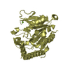
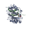
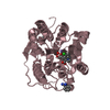
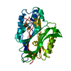
| Title | HUMAN S-ADENOSYLMETHIONINE DECARBOXYLASE | |||||||||
 Components Components |
| |||||||||
 Keywords Keywords | S-ADENOSYLMETHIONINE DECARBOXYLASE / PYRUVOYL / GENE DUPLICATION / POLYAMINE BIOSYNTHESIS / SANDWICH / ALLOSTERIC ENZYME | |||||||||
| Function / homology |  Function and homology information Function and homology informationspermine biosynthetic process / adenosylmethionine decarboxylase / adenosylmethionine decarboxylase activity / Metabolism of polyamines / polyamine metabolic process / putrescine binding / spermidine biosynthetic process / identical protein binding / cytosol Similarity search - Function | |||||||||
| Biological species |  Homo sapiens (human) Homo sapiens (human) | |||||||||
| Method |  X-RAY DIFFRACTION / SYNCHROTRON / MAD / Resolution: 2.25 Å X-RAY DIFFRACTION / SYNCHROTRON / MAD / Resolution: 2.25 Å | |||||||||
 Authors Authors | Ekstrom, J.L. / Mathews, I.I. / Stanley, B.A. / Pegg, A.E. / Ealick, S.E. | |||||||||
 Citation Citation | Journal: Structure Fold.Des. / Year: 1999 Title: The crystal structure of human S-adenosylmethionine decarboxylase at 2.25 A resolution reveals a novel fold. Authors: Ekstrom, J.L. / Mathews, I.I. / Stanley, B.A. / Pegg, A.E. / Ealick, S.E. | |||||||||
| History |
|
- Structure visualization
| Structure viewer | Molecule: MolmilJmol/JSmol |
|---|
- Downloads & links
-Download
| PDBx/mmCIF format | 1jen.cif.gz | 141.7 KB | Display | PDBx/mmCIF format |
|---|---|---|---|---|
| PDB format | pdb1jen.ent.gz | 110.4 KB | Display | PDB format |
| PDBx/mmJSON format | 1jen.json.gz | Tree view | PDBx/mmJSON format | |
| Others |  Other downloads Other downloads |
-Validation report
| Arichive directory | https://data.pdbj.org/pub/pdb/validation_reports/je/1jenftp://data.pdbj.org/pub/pdb/validation_reports/je/1jen | HTTPS FTP |
|---|
-Related structure data
| Similar structure data |
|---|


-Links
 PDBj
PDBj- Assembly
| Deposited unit | 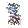
| ||||||||
|---|---|---|---|---|---|---|---|---|---|
| 1 | 
| ||||||||
| 2 | 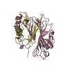
| ||||||||
| Unit cell |
| ||||||||
| Noncrystallographic symmetry (NCS) | NCS oper: (Code: given Matrix: (-0.998116, 0.057715, 0.020825), Vector: |
-Components
| #1: Protein | Mass: 7694.577 Da / Num. of mol.: 2 Source method: isolated from a genetically manipulated source Source: (gene. exp.) Homo sapiens (human) / Production host:  References: UniProt: P17707, adenosylmethionine decarboxylase #2: Protein | Mass: 30772.105 Da / Num. of mol.: 2 Source method: isolated from a genetically manipulated source Source: (gene. exp.) Homo sapiens (human) / Production host: References: UniProt: P17707, adenosylmethionine decarboxylase #3: Water | ChemComp-HOH / |  Mass: 18.015 Da / Num. of mol.: 414 / Source method: isolated from a natural source / Formula: H2O Mass: 18.015 Da / Num. of mol.: 414 / Source method: isolated from a natural source / Formula: H2OHas protein modification | Y | |
|---|
-Experimental details
-Experiment
| Experiment | Method: X-RAY DIFFRACTION / Number of used crystals: 1 |
|---|
- Sample preparation
| Crystal | Density Matthews: 2.3 Å3/Da / Density % sol: 49 % / Description: 22 SELENIUM ATOMS LOCATED IN ASYMMETRIC UNIT | |||||||||||||||
|---|---|---|---|---|---|---|---|---|---|---|---|---|---|---|---|---|
| Crystal grow | pH: 8 / Details: 12 - 16% PEG 8K, 10 MM TRIS-HCL, PH 8.0, pH 8.00 | |||||||||||||||
| Crystal | *PLUS Density % sol: 48 % | |||||||||||||||
| Crystal grow | *PLUS Method: vapor diffusion, hanging drop | |||||||||||||||
| Components of the solutions | *PLUS
|
-Data collection
| Diffraction | Mean temperature: 93 K | ||||||||||||
|---|---|---|---|---|---|---|---|---|---|---|---|---|---|
| Diffraction source | Source: SYNCHROTRON / Site: NSLS  / Beamline: X12C / Wavelength: 0.9791,0.9788,0.954 / Beamline: X12C / Wavelength: 0.9791,0.9788,0.954 | ||||||||||||
| Detector | Type: BRANDEIS / Detector: CCD / Date: Sep 1, 1997 | ||||||||||||
| Radiation | Protocol: MAD / Monochromatic (M) / Laue (L): M / Scattering type: x-ray | ||||||||||||
| Radiation wavelength |
| ||||||||||||
| Reflection | Resolution: 2.25→20 Å / Num. obs: 169725 / % possible obs: 98.9 % / Observed criterion σ(I): 2 / Redundancy: 5.1 % / Biso Wilson estimate: 15.95 Å2 / Rsym value: 0.05 / Net I/σ(I): 23.71 | ||||||||||||
| Reflection shell | Resolution: 2.25→2.33 Å / Redundancy: 2.8 % / Mean I/σ(I) obs: 15 / Rsym value: 0.094 / % possible all: 98.1 | ||||||||||||
| Reflection | *PLUS Num. obs: 33426 / % possible obs: 98.9 % / Num. measured all: 169725 / Rmerge(I) obs: 0.05 | ||||||||||||
| Reflection shell | *PLUS % possible obs: 98.1 % / Rmerge(I) obs: 0.094 |
- Processing
| Software |
| ||||||||||||||||||||||||||||||||||||||||||||||||||||||||||||
|---|---|---|---|---|---|---|---|---|---|---|---|---|---|---|---|---|---|---|---|---|---|---|---|---|---|---|---|---|---|---|---|---|---|---|---|---|---|---|---|---|---|---|---|---|---|---|---|---|---|---|---|---|---|---|---|---|---|---|---|---|---|
| Refinement | Method to determine structure: MAD / Resolution: 2.25→20 Å / Rfactor Rfree error: 0.005 / Data cutoff high absF: 1000000 / Data cutoff low absF: 0.001 / Cross valid method: THROUGHOUT / σ(F): 2
| ||||||||||||||||||||||||||||||||||||||||||||||||||||||||||||
| Displacement parameters | Biso mean: 20.45 Å2 | ||||||||||||||||||||||||||||||||||||||||||||||||||||||||||||
| Refine analyze |
| ||||||||||||||||||||||||||||||||||||||||||||||||||||||||||||
| Refinement step | Cycle: LAST / Resolution: 2.25→20 Å
| ||||||||||||||||||||||||||||||||||||||||||||||||||||||||||||
| Refine LS restraints |
| ||||||||||||||||||||||||||||||||||||||||||||||||||||||||||||
| Refine LS restraints NCS | NCS model details: RESTRAINTS | ||||||||||||||||||||||||||||||||||||||||||||||||||||||||||||
| LS refinement shell | Resolution: 2.25→2.35 Å / Rfactor Rfree error: 0.02 / Total num. of bins used: 8
| ||||||||||||||||||||||||||||||||||||||||||||||||||||||||||||
| Software | *PLUS Name: X-PLOR / Version: 3.8 / Classification: refinement | ||||||||||||||||||||||||||||||||||||||||||||||||||||||||||||
| Refinement | *PLUS Rfactor obs: 0.177 | ||||||||||||||||||||||||||||||||||||||||||||||||||||||||||||
| Solvent computation | *PLUS | ||||||||||||||||||||||||||||||||||||||||||||||||||||||||||||
| Displacement parameters | *PLUS | ||||||||||||||||||||||||||||||||||||||||||||||||||||||||||||
| Refine LS restraints | *PLUS
| ||||||||||||||||||||||||||||||||||||||||||||||||||||||||||||
| LS refinement shell | *PLUS Rfactor obs: 0.2255 |
