 Movie
Movie Controller

[English] 日本語
 Yorodumi
- PDB-1vcu: Structure of the human cytosolic sialidase Neu2 in complex with t... -
+ Open data

- Basic information
| Entry | Database: PDB / ID: 1vcu | ||||||
|---|---|---|---|---|---|---|---|
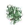
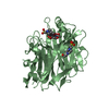
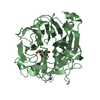
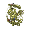
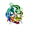
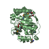
| Title | Structure of the human cytosolic sialidase Neu2 in complex with the inhibitor DANA | ||||||
 Components Components | Sialidase 2 | ||||||
 Keywords Keywords | HYDROLASE / sialidase / neuraminidase / ganglioside / dana / sialic acid | ||||||
| Function / homology |  Function and homology information Function and homology informationSialic acid metabolism / glycosphingolipid catabolic process / glycoprotein catabolic process / Glycosphingolipid catabolism / ganglioside catabolic process / oligosaccharide catabolic process / exo-alpha-sialidase / exo-alpha-sialidase activity / catalytic complex / lysosome ...Sialic acid metabolism / glycosphingolipid catabolic process / glycoprotein catabolic process / Glycosphingolipid catabolism / ganglioside catabolic process / oligosaccharide catabolic process / exo-alpha-sialidase / exo-alpha-sialidase activity / catalytic complex / lysosome / membrane / cytoplasm / cytosol Similarity search - Function | ||||||
| Biological species |  Homo sapiens (human) Homo sapiens (human) | ||||||
| Method |  X-RAY DIFFRACTION / SYNCHROTRON / MOLECULAR REPLACEMENT / Resolution: 2.85 Å X-RAY DIFFRACTION / SYNCHROTRON / MOLECULAR REPLACEMENT / Resolution: 2.85 Å | ||||||
 Authors Authors | Chavas, L.M.G. / Fusi, P. / Tringali, C. / Venerando, B. / Tettamanti, G. / Kato, R. / Monti, E. / Wakatsuki, S. | ||||||
 Citation Citation | Journal: J.Biol.Chem. / Year: 2005 Title: Crystal Structure of the Human Cytosolic Sialidase Neu2: EVIDENCE FOR THE DYNAMIC NATURE OF SUBSTRATE RECOGNITION Authors: Chavas, L.M.G. / Tringali, C. / Fusi, P. / Venerando, B. / Tettamanti, G. / Kato, R. / Monti, E. / Wakatsuki, S. | ||||||
| History |
|
- Structure visualization
| Structure viewer | Molecule: MolmilJmol/JSmol |
|---|
- Downloads & links
-Download
| PDBx/mmCIF format | 1vcu.cif.gz | 158.8 KB | Display | PDBx/mmCIF format |
|---|---|---|---|---|
| PDB format | pdb1vcu.ent.gz | 125.1 KB | Display | PDB format |
| PDBx/mmJSON format | 1vcu.json.gz | Tree view | PDBx/mmJSON format | |
| Others |  Other downloads Other downloads |
-Validation report
| Arichive directory | https://data.pdbj.org/pub/pdb/validation_reports/vc/1vcuftp://data.pdbj.org/pub/pdb/validation_reports/vc/1vcu | HTTPS FTP |
|---|
-Related structure data
| Related structure data |  1sntSC  1so7C S: Starting model for refinement C: citing same article ( |
|---|---|
| Similar structure data |


-Links
 PDBj
PDBj

- Assembly
| Deposited unit | 
| ||||||||
|---|---|---|---|---|---|---|---|---|---|
| 1 | 
| ||||||||
| 2 | 
| ||||||||
| Unit cell |
|
-Components
| #1: Protein | Mass: 42419.598 Da / Num. of mol.: 2 Source method: isolated from a genetically manipulated source Source: (gene. exp.) Homo sapiens (human) / Plasmid: pGEX-2T / Species (production host): Escherichia coli / Production host:  #2: Sugar |   Type: D-saccharide / Mass: 291.255 Da / Num. of mol.: 2 / Source method: obtained synthetically / Formula: C11H17NO8 Type: D-saccharide / Mass: 291.255 Da / Num. of mol.: 2 / Source method: obtained synthetically / Formula: C11H17NO8#3: Chemical | ChemComp-EPE /   Mass: 238.305 Da / Num. of mol.: 4 / Source method: obtained synthetically / Formula: C8H18N2O4S / Comment: pH buffer*YM Mass: 238.305 Da / Num. of mol.: 4 / Source method: obtained synthetically / Formula: C8H18N2O4S / Comment: pH buffer*YM#4: Water | ChemComp-HOH / |  Mass: 18.015 Da / Num. of mol.: 131 / Source method: isolated from a natural source / Formula: H2O Mass: 18.015 Da / Num. of mol.: 131 / Source method: isolated from a natural source / Formula: H2O |
|---|
-Experimental details
-Experiment
| Experiment | Method: X-RAY DIFFRACTION / Number of used crystals: 1 |
|---|
- Sample preparation
| Crystal | Density Matthews: 2.64 Å3/Da / Density % sol: 53.12 % |
|---|---|
| Crystal grow | Temperature: 289 K / Method: vapor diffusion, hanging drop / pH: 7 Details: 2-deoxy-2,3-dehydro-N-acetyl neuraminic acid, 2-methyl-2,4-pentanediol, guanidine hydrochloride, pH 7.0, VAPOR DIFFUSION, HANGING DROP, temperature 289K |
-Data collection
| Diffraction | Mean temperature: 100 K |
|---|---|
| Diffraction source | Source: SYNCHROTRON / Site: Photon Factory  / Beamline: BL-6A / Wavelength: 0.97 Å / Beamline: BL-6A / Wavelength: 0.97 Å |
| Detector | Type: ADSC QUAMTUM 4r / Detector: CCD / Date: Jun 27, 2003 |
| Radiation | Monochromator: Si(111) / Protocol: SINGLE WAVELENGTH / Monochromatic (M) / Laue (L): M / Scattering type: x-ray |
| Radiation wavelength | Wavelength: 0.97 Å / Relative weight: 1 |
| Reflection | Resolution: 2.85→50 Å / Num. all: 20889 / Num. obs: 20807 / % possible obs: 99.7 % / Rmerge(I) obs: 0.115 / Net I/σ(I): 11.5 |
| Reflection shell | Resolution: 2.85→2.95 Å / Rmerge(I) obs: 0.313 / Mean I/σ(I) obs: 2 / % possible all: 99.6 |
- Processing
| Software |
| |||||||||||||||||||||||||
|---|---|---|---|---|---|---|---|---|---|---|---|---|---|---|---|---|---|---|---|---|---|---|---|---|---|---|
| Refinement | Method to determine structure: MOLECULAR REPLACEMENT Starting model: PDB ENTRY 1SNT Resolution: 2.85→50 Å / Cross valid method: THROUGHOUT / σ(F): 0 / Stereochemistry target values: Engh & Huber
| |||||||||||||||||||||||||
| Refinement step | Cycle: LAST / Resolution: 2.85→50 Å
| |||||||||||||||||||||||||
| Refine LS restraints |
|
