 Movie
Movie Controller


+ Open data

- Basic information
| Entry | Database: PDB / ID: 1f76 | ||||||
|---|---|---|---|---|---|---|---|
| Title | ESCHERICHIA COLI DIHYDROOROTATE DEHYDROGENASE | ||||||
 Components Components | Dihydroorotate dehydrogenase (quinone) | ||||||
 Keywords Keywords | OXIDOREDUCTASE / MONOMER / ALPHA-BETA-BARREL / FMN BINDING DOMAIN / OROTATE COMPLEX | ||||||
| Function / homology |  Function and homology information Function and homology informationpyrimidine ribonucleotide biosynthetic process / dihydroorotate dehydrogenase (quinone) / dihydroorotate dehydrogenase (quinone) activity / dihydroorotate dehydrogenase activity / 'de novo' UMP biosynthetic process / 'de novo' pyrimidine nucleobase biosynthetic process / FMN binding / membrane / plasma membrane / cytosol Similarity search - Function | ||||||
| Biological species |  | ||||||
| Method |  X-RAY DIFFRACTION / SYNCHROTRON / MAD / Resolution: 2.5 Å X-RAY DIFFRACTION / SYNCHROTRON / MAD / Resolution: 2.5 Å | ||||||
 Authors Authors | Norager, S. / Jensen, K.F. / Bjornberg, O. / Larsen, S. | ||||||
 Citation Citation | Journal: Structure / Year: 2002 Title: E. coli Dihydroorotate Dehydrogenase Reveals Structural and Functional Distinction between different classes of dihydroorotate dehydrogenases Authors: Norager, S. / Jensen, K.F. / Bjornberg, O. / Larsen, S. #1: Journal: Acta Crystallogr.,Sect.D / Year: 2000Title: Crystallization and Preliminary X-ray Studies of Membrane-associated Escherichia coli Dihydroorotate Dehydrogenase. Authors: Rowland, P. / Norager, S. / Jensen, K.F. / Larsen, S. #2: Journal: Biochemistry / Year: 1999Title: The Activity of Escherichia coli Dihydroorotate Dehydrogenase is Dependent on a Conserved Loop Identified by Sequence Homology, Mutagenesis and Limited Proteolysis. Authors: Bjornberg, O. / Gruner, A.-C. / Roepstorff, P. / Jensen, K.F. #3: Journal: Flavins and Flavoproteins,Proc.13th Int.Symp. / Year: 1999Title: The Dihydroorotate Dehydrogenases of Escherichia coli and Lactococcus Lactis Represent Two Distinct Families of the Enzyme. Authors: Bjornberg, O. / Jensen, K.F. / Gruner, A.-C. / Ottosen, M. / Sorensen, P. / Rowland, P. / Norager, S. / Larsen, S. #4: Journal: Flavins and Flavoproteins,Proc.13th Int.Symp. / Year: 1999Title: Reduction Reactions of Two Dihydroorotate Dehydrogenases. Authors: Palfey, B. / Bjornberg, O. / Jensen, K.F. | ||||||
| History |
|
- Structure visualization
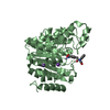
| Structure viewer | Molecule: MolmilJmol/JSmol |
|---|
- Downloads & links
-Download
| PDBx/mmCIF format | 1f76.cif.gz | 296.3 KB | Display | PDBx/mmCIF format |
|---|---|---|---|---|
| PDB format | pdb1f76.ent.gz | 239.3 KB | Display | PDB format |
| PDBx/mmJSON format | 1f76.json.gz | Tree view | PDBx/mmJSON format | |
| Others |  Other downloads Other downloads |
-Validation report
| Arichive directory | https://data.pdbj.org/pub/pdb/validation_reports/f7/1f76ftp://data.pdbj.org/pub/pdb/validation_reports/f7/1f76 | HTTPS FTP |
|---|
-Related structure data
| Similar structure data |
|---|


-Links
 PDBj
PDBj- Assembly
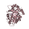
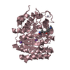
| Deposited unit | 
| ||||||||
|---|---|---|---|---|---|---|---|---|---|
| 1 | 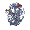
| ||||||||
| 2 | 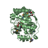
| ||||||||
| 3 | 
| ||||||||
| 4 | 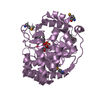
| ||||||||
| Unit cell |
| ||||||||
| Details | The biological assembly is a monomer. The Biological Assembly is a Monomer. |
-Components
| #1: Protein | Mass: 37146.711 Da / Num. of mol.: 4 Source method: isolated from a genetically manipulated source Source: (gene. exp.) References: UniProt: P0A7E1, dihydroorotate dehydrogenase (quinone) #2: Chemical | ChemComp-FMN /   Mass: 456.344 Da / Num. of mol.: 4 / Source method: obtained synthetically / Formula: C17H21N4O9P Mass: 456.344 Da / Num. of mol.: 4 / Source method: obtained synthetically / Formula: C17H21N4O9P#3: Chemical | 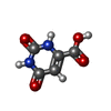  Mass: 156.096 Da / Num. of mol.: 3 / Source method: obtained synthetically / Formula: C5H4N2O4 Mass: 156.096 Da / Num. of mol.: 3 / Source method: obtained synthetically / Formula: C5H4N2O4#4: Chemical | ChemComp-FMT /   Mass: 46.025 Da / Num. of mol.: 8 / Source method: obtained synthetically / Formula: CH2O2 Mass: 46.025 Da / Num. of mol.: 8 / Source method: obtained synthetically / Formula: CH2O2#5: Water | ChemComp-HOH / |  Mass: 18.015 Da / Num. of mol.: 1043 / Source method: isolated from a natural source / Formula: H2O Mass: 18.015 Da / Num. of mol.: 1043 / Source method: isolated from a natural source / Formula: H2OHas protein modification | Y | |
|---|
-Experimental details
-Experiment
| Experiment | Method: X-RAY DIFFRACTION / Number of used crystals: 1 |
|---|
- Sample preparation
| Crystal | Density Matthews: 3.57 Å3/Da / Density % sol: 65.5 % | |||||||||||||||||||||||||||||||||||||||||||||||||||||||||||||||
|---|---|---|---|---|---|---|---|---|---|---|---|---|---|---|---|---|---|---|---|---|---|---|---|---|---|---|---|---|---|---|---|---|---|---|---|---|---|---|---|---|---|---|---|---|---|---|---|---|---|---|---|---|---|---|---|---|---|---|---|---|---|---|---|---|
| Crystal grow | Temperature: 298 K / Method: vapor diffusion, sitting drop / pH: 3.5 Details: 3.3 to 3.7 M Na-formate, 0.1 M Na-acetate, 25 mM b-OG, 12-15 mg/ml protein, pH 3.5, VAPOR DIFFUSION, SITTING DROP, temperature 298K | |||||||||||||||||||||||||||||||||||||||||||||||||||||||||||||||
| Crystal grow | *PLUS pH: 7 | |||||||||||||||||||||||||||||||||||||||||||||||||||||||||||||||
| Components of the solutions | *PLUS
|
-Data collection
| Diffraction | Mean temperature: 108 K |
|---|---|
| Diffraction source | Source: SYNCHROTRON / Site: ESRF  / Beamline: BM30A / Wavelength: 0.97 / Beamline: BM30A / Wavelength: 0.97 |
| Detector | Type: MARRESEARCH / Detector: IMAGE PLATE / Date: Jun 25, 1999 |
| Radiation | Protocol: MULTIPLE ANOMALOUS DIFFRACTION / Monochromatic (M) / Laue (L): M / Scattering type: x-ray |
| Radiation wavelength | Wavelength: 0.97 Å / Relative weight: 1 |
| Reflection | Resolution: 2.5→50 Å / Num. all: 75583 / Num. obs: 920967 / % possible obs: 98 % / Observed criterion σ(F): 0 / Observed criterion σ(I): 0 / Redundancy: 8.2 % / Biso Wilson estimate: 34.2 Å2 / Rmerge(I) obs: 0.085 / Net I/σ(I): 16.1 |
| Reflection shell | Resolution: 2.5→2.54 Å / Rmerge(I) obs: 0.334 / Num. unique all: 3137 / % possible all: 84.6 |
| Reflection | *PLUS Num. obs: 75583 / % possible obs: 98 % / Rmerge(I) obs: 0.085 |
| Reflection shell | *PLUS % possible obs: 84.6 % / Rmerge(I) obs: 0.034 / Mean I/σ(I) obs: 3.5 |
- Processing
| Software |
| ||||||||||||||||||||||||||||||||||||||||||||||||||||||||||||||||||||||||||||||||
|---|---|---|---|---|---|---|---|---|---|---|---|---|---|---|---|---|---|---|---|---|---|---|---|---|---|---|---|---|---|---|---|---|---|---|---|---|---|---|---|---|---|---|---|---|---|---|---|---|---|---|---|---|---|---|---|---|---|---|---|---|---|---|---|---|---|---|---|---|---|---|---|---|---|---|---|---|---|---|---|---|---|
| Refinement | Method to determine structure: MAD / Resolution: 2.5→20 Å / Rfactor Rfree error: 0.003 / Isotropic thermal model: RESTRAINED / Cross valid method: THROUGHOUT / σ(F): 0 / σ(I): 0 / Stereochemistry target values: Engh & Huber Details: Structure Solved from a MAD Data Set Collected on the Se-methionine Substituted Enzyme
| ||||||||||||||||||||||||||||||||||||||||||||||||||||||||||||||||||||||||||||||||
| Solvent computation | Solvent model: FLAT MODEL / Bsol: 44.1302 Å2 / ksol: 0.336959 e/Å3 | ||||||||||||||||||||||||||||||||||||||||||||||||||||||||||||||||||||||||||||||||
| Displacement parameters | Biso mean: 32.2 Å2
| ||||||||||||||||||||||||||||||||||||||||||||||||||||||||||||||||||||||||||||||||
| Refine analyze |
| ||||||||||||||||||||||||||||||||||||||||||||||||||||||||||||||||||||||||||||||||
| Refinement step | Cycle: LAST / Resolution: 2.5→20 Å
| ||||||||||||||||||||||||||||||||||||||||||||||||||||||||||||||||||||||||||||||||
| Refine LS restraints |
| ||||||||||||||||||||||||||||||||||||||||||||||||||||||||||||||||||||||||||||||||
| LS refinement shell | Resolution: 2.5→2.66 Å / Rfactor Rfree error: 0.008 / Total num. of bins used: 6
| ||||||||||||||||||||||||||||||||||||||||||||||||||||||||||||||||||||||||||||||||
| Refinement | *PLUS Rfactor Rfree: 0.217 / Rfactor Rwork: 0.177 | ||||||||||||||||||||||||||||||||||||||||||||||||||||||||||||||||||||||||||||||||
| Solvent computation | *PLUS | ||||||||||||||||||||||||||||||||||||||||||||||||||||||||||||||||||||||||||||||||
| Displacement parameters | *PLUS | ||||||||||||||||||||||||||||||||||||||||||||||||||||||||||||||||||||||||||||||||
| Refine LS restraints | *PLUS
| ||||||||||||||||||||||||||||||||||||||||||||||||||||||||||||||||||||||||||||||||
| LS refinement shell | *PLUS Rfactor Rfree: 0.271 / Rfactor Rwork: 0.219 |
