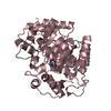
 Movie
Movie Controller


+ Open data

- Basic information
| Entry | Database: PDB / ID: 1a4m | ||||||
|---|---|---|---|---|---|---|---|
| Title | ADA STRUCTURE COMPLEXED WITH PURINE RIBOSIDE AT PH 7.0 | ||||||
 Components Components | ADENOSINE DEAMINASE | ||||||
 Keywords Keywords | HYDROLASE / ADENOSINE DEAMINASE / PURINE RIBOSIDE | ||||||
| Function / homology |  Function and homology information Function and homology informationxanthine biosynthetic process / negative regulation of penile erection / Purine salvage / Ribavirin ADME / purine nucleotide salvage / negative regulation of circadian sleep/wake cycle, non-REM sleep / negative regulation of mucus secretion / purine nucleoside binding / positive regulation of germinal center formation / 2'-deoxyadenosine deaminase activity ...xanthine biosynthetic process / negative regulation of penile erection / Purine salvage / Ribavirin ADME / purine nucleotide salvage / negative regulation of circadian sleep/wake cycle, non-REM sleep / negative regulation of mucus secretion / purine nucleoside binding / positive regulation of germinal center formation / 2'-deoxyadenosine deaminase activity / negative regulation of adenosine receptor signaling pathway / cytoplasmic vesicle lumen / hypoxanthine biosynthetic process / histamine secretion / deoxyadenosine catabolic process / dAMP catabolic process / inhibition of non-skeletal tissue mineralization / germinal center B cell differentiation / deaminase activity / positive regulation of T cell differentiation in thymus / AMP catabolic process / adenosine deaminase / inosine biosynthetic process / dATP catabolic process / adenosine metabolic process / negative regulation of leukocyte migration / hypoxanthine salvage / adenosine deaminase activity / adenosine catabolic process / regulation of circadian sleep/wake cycle, sleep / trophectodermal cell differentiation / regulation of cell-cell adhesion mediated by integrin / embryonic digestive tract development / IMP salvage / regulation of T cell differentiation in thymus / Peyer's patch development / positive regulation of smooth muscle contraction / negative regulation of mature B cell apoptotic process / positive regulation of alpha-beta T cell differentiation / response to purine-containing compound / negative regulation of thymocyte apoptotic process / positive regulation of T cell receptor signaling pathway / anchoring junction / lung alveolus development / regulation of T cell differentiation / positive regulation of T cell differentiation / response to vitamin E / positive regulation of heart rate / positive regulation of B cell proliferation / positive regulation of calcium-mediated signaling / lung development / placenta development / dendrite cytoplasm / liver development / T cell activation / negative regulation of inflammatory response / positive regulation of T cell activation / in utero embryonic development / response to hypoxia / lysosome / cell adhesion / external side of plasma membrane / neuronal cell body / negative regulation of apoptotic process / cell surface / : / zinc ion binding / membrane / plasma membrane / cytosol / cytoplasm Similarity search - Function | ||||||
| Biological species |  | ||||||
| Method |  X-RAY DIFFRACTION / MOLECULAR REPLACEMENT / Resolution: 1.95 Å X-RAY DIFFRACTION / MOLECULAR REPLACEMENT / Resolution: 1.95 Å | ||||||
 Authors Authors | Wang, Z. / Quiocho, F.A. | ||||||
 Citation Citation | Journal: Biochemistry / Year: 1998 Title: Complexes of adenosine deaminase with two potent inhibitors: X-ray structures in four independent molecules at pH of maximum activity. Authors: Wang, Z. / Quiocho, F.A. #1: Journal: Biochemistry / Year: 1993Title: A Pre-Transition-State Mimic of an Enzyme: X-Ray Structure of Adenosine Deaminase with Bound 1-Deazaadenosine and Zinc-Activated Water Authors: Wilson, D.K. / Quiocho, F.A. #2: Journal: J.Mol.Biol. / Year: 1992Title: Refined 2.5 A Structure of Murine Adenosine Deaminase at Ph 6.0 Authors: Sharff, A.J. / Wilson, D.K. / Chang, Z. / Quiocho, F.A. #3: Journal: Science / Year: 1991Title: Atomic Structure of Adenosine Deaminase Complexed with a Transition-State Analog: Understanding Catalysis and Immunodeficiency Mutations Authors: Wilson, D.K. / Rudolph, F.B. / Quiocho, F.A. | ||||||
| History |
|
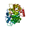
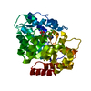
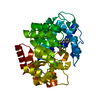
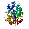
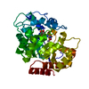
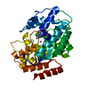
- Structure visualization
| Structure viewer | Molecule: MolmilJmol/JSmol |
|---|
- Downloads & links
-Download
| PDBx/mmCIF format | 1a4m.cif.gz | 314.5 KB | Display | PDBx/mmCIF format |
|---|---|---|---|---|
| PDB format | pdb1a4m.ent.gz | 248.1 KB | Display | PDB format |
| PDBx/mmJSON format | 1a4m.json.gz | Tree view | PDBx/mmJSON format | |
| Others |  Other downloads Other downloads |
-Validation report
| Arichive directory | https://data.pdbj.org/pub/pdb/validation_reports/a4/1a4mftp://data.pdbj.org/pub/pdb/validation_reports/a4/1a4m | HTTPS FTP |
|---|
-Related structure data
| Related structure data |  1a4lC  2adaS S: Starting model for refinement C: citing same article ( |
|---|---|
| Similar structure data |


-Links
 PDBj
PDBj

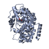
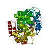
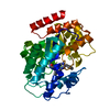
- Assembly
| Deposited unit | 
| ||||||||
|---|---|---|---|---|---|---|---|---|---|
| 1 | 
| ||||||||
| 2 | 
| ||||||||
| 3 | 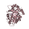
| ||||||||
| 4 | 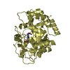
| ||||||||
| Unit cell |
|
-Components
| #1: Protein | Mass: 39715.188 Da / Num. of mol.: 4 Source method: isolated from a genetically manipulated source Source: (gene. exp.)  #2: Chemical | ChemComp-ZN /   Mass: 65.409 Da / Num. of mol.: 4 / Source method: obtained synthetically / Formula: Zn Mass: 65.409 Da / Num. of mol.: 4 / Source method: obtained synthetically / Formula: Zn#3: Chemical | ChemComp-PRH / 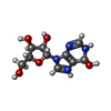  Mass: 271.250 Da / Num. of mol.: 4 / Source method: obtained synthetically / Formula: C10H15N4O5 Mass: 271.250 Da / Num. of mol.: 4 / Source method: obtained synthetically / Formula: C10H15N4O5#4: Water | ChemComp-HOH / |  Mass: 18.015 Da / Num. of mol.: 1065 / Source method: isolated from a natural source / Formula: H2O Mass: 18.015 Da / Num. of mol.: 1065 / Source method: isolated from a natural source / Formula: H2O |
|---|
-Experimental details
-Experiment
| Experiment | Method: X-RAY DIFFRACTION / Number of used crystals: 1 |
|---|
- Sample preparation
| Crystal | Density Matthews: 2.6 Å3/Da / Density % sol: 58 % | ||||||||||||||||||||||||||||||||||||||||||||||||||||||
|---|---|---|---|---|---|---|---|---|---|---|---|---|---|---|---|---|---|---|---|---|---|---|---|---|---|---|---|---|---|---|---|---|---|---|---|---|---|---|---|---|---|---|---|---|---|---|---|---|---|---|---|---|---|---|---|
| Crystal grow | pH: 7 / Details: 20% PEG 3350, 100 MM NACL, 100 MM HEPES PH 7.0 | ||||||||||||||||||||||||||||||||||||||||||||||||||||||
| Crystal | *PLUS Density % sol: 65 % | ||||||||||||||||||||||||||||||||||||||||||||||||||||||
| Crystal grow | *PLUS Temperature: 4 ℃ / Method: vapor diffusion, hanging drop | ||||||||||||||||||||||||||||||||||||||||||||||||||||||
| Components of the solutions | *PLUS
|
-Data collection
| Diffraction | Mean temperature: 100 K |
|---|---|
| Diffraction source | Source: ROTATING ANODE / Type: RIGAKU RUH2R / Wavelength: 1.5418 |
| Detector | Type: MACSCIENCE / Detector: IMAGE PLATE / Date: May 15, 1997 / Details: MIRROR |
| Radiation | Monochromator: NI FILTER / Monochromatic (M) / Laue (L): M / Scattering type: x-ray |
| Radiation wavelength | Wavelength: 1.5418 Å / Relative weight: 1 |
| Reflection | Resolution: 1.95→10 Å / Num. obs: 83321 / % possible obs: 70.2 % / Observed criterion σ(I): 0 / Redundancy: 4 % / Rsym value: 0.078 / Net I/σ(I): 8 |
| Reflection shell | Resolution: 1.95→2.02 Å / Redundancy: 1.9 % / Mean I/σ(I) obs: 2.1 / Rsym value: 0.401 / % possible all: 47 |
| Reflection | *PLUS Num. measured all: 415190 / Rmerge(I) obs: 0.078 |
| Reflection shell | *PLUS % possible obs: 47 % / Rmerge(I) obs: 0.401 |
- Processing
| Software |
| ||||||||||||||||||||||||||||||||||||||||||||||||||||||||||||
|---|---|---|---|---|---|---|---|---|---|---|---|---|---|---|---|---|---|---|---|---|---|---|---|---|---|---|---|---|---|---|---|---|---|---|---|---|---|---|---|---|---|---|---|---|---|---|---|---|---|---|---|---|---|---|---|---|---|---|---|---|---|
| Refinement | Method to determine structure: MOLECULAR REPLACEMENT Starting model: PDB ENTRY 2ADA Resolution: 1.95→10 Å / Rfactor Rfree error: 0.003 / Data cutoff high absF: 10000000 / Data cutoff low absF: 0 / Cross valid method: THROUGHOUT / σ(F): 0
| ||||||||||||||||||||||||||||||||||||||||||||||||||||||||||||
| Displacement parameters | Biso mean: 16.96 Å2 | ||||||||||||||||||||||||||||||||||||||||||||||||||||||||||||
| Refinement step | Cycle: LAST / Resolution: 1.95→10 Å
| ||||||||||||||||||||||||||||||||||||||||||||||||||||||||||||
| Refine LS restraints |
| ||||||||||||||||||||||||||||||||||||||||||||||||||||||||||||
| Software | *PLUS Name: X-PLOR / Version: 3.85 / Classification: refinement | ||||||||||||||||||||||||||||||||||||||||||||||||||||||||||||
| Refinement | *PLUS Num. reflection obs: 77948 | ||||||||||||||||||||||||||||||||||||||||||||||||||||||||||||
| Solvent computation | *PLUS | ||||||||||||||||||||||||||||||||||||||||||||||||||||||||||||
| Displacement parameters | *PLUS | ||||||||||||||||||||||||||||||||||||||||||||||||||||||||||||
| Refine LS restraints | *PLUS
|
