 Movie
Movie Controller


+ Open data

- Basic information
| Entry | Database: PDB / ID: 1jef | |||||||||
|---|---|---|---|---|---|---|---|---|---|---|
| Title | TURKEY LYSOZYME COMPLEX WITH (GLCNAC)3 | |||||||||
 Components Components | LYSOZYME | |||||||||
 Keywords Keywords | HYDROLASE / ENZYME / INHIBITOR COMPLEX / GLYCOSIDASE / BACTERIOLYTIC ENZYME | |||||||||
| Function / homology |  Function and homology information Function and homology informationglycosaminoglycan binding / cell wall macromolecule catabolic process / lysozyme / lysozyme activity / killing of cells of another organism / defense response to Gram-negative bacterium / defense response to Gram-positive bacterium / : / identical protein binding / cytoplasm Similarity search - Function | |||||||||
| Biological species |  | |||||||||
| Method |  X-RAY DIFFRACTION / MOLECULAR REPLACEMENT / Resolution: 1.77 Å X-RAY DIFFRACTION / MOLECULAR REPLACEMENT / Resolution: 1.77 Å | |||||||||
 Authors Authors | Harata, K. / Muraki, M. | |||||||||
 Citation Citation | Journal: Acta Crystallogr.,Sect.D / Year: 1997 Title: X-ray structure of turkey-egg lysozyme complex with tri-N-acetylchitotriose. Lack of binding ability at subsite A. Authors: Harata, K. / Muraki, M. #1: Journal: Acta Crystallogr.,Sect.D / Year: 1995Title: X-Ray Structure of Turkey Egg Lysozyme Complex with Di-N-Acetylchitobiose. Recognition and Binding of Alpha-Anomeric Form Authors: Harata, K. / Muraki, M. #2: Journal: Acta Crystallogr.,Sect.D / Year: 1993Title: X-Ray Structure of Monoclinic Turkey Egg Lysozyme at 1.3 Angstrom Resolution Authors: Harata, K. | |||||||||
| History |
|
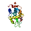
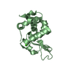
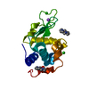
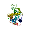
- Structure visualization
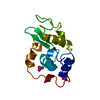
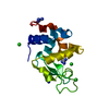
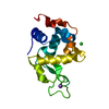
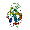
| Structure viewer | Molecule: MolmilJmol/JSmol |
|---|
- Downloads & links
-Download
| PDBx/mmCIF format | 1jef.cif.gz | 42.3 KB | Display | PDBx/mmCIF format |
|---|---|---|---|---|
| PDB format | pdb1jef.ent.gz | 28.3 KB | Display | PDB format |
| PDBx/mmJSON format | 1jef.json.gz | Tree view | PDBx/mmJSON format | |
| Others |  Other downloads Other downloads |
-Validation report
| Arichive directory | https://data.pdbj.org/pub/pdb/validation_reports/je/1jefftp://data.pdbj.org/pub/pdb/validation_reports/je/1jef | HTTPS FTP |
|---|
-Related structure data
| Similar structure data |
|---|
-Links
 PDBj
PDBj
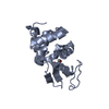
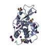
- Assembly
| Deposited unit | 
| ||||||||
|---|---|---|---|---|---|---|---|---|---|
| 1 |
| ||||||||
| Unit cell |
| ||||||||
| Components on special symmetry positions |
|
-Components
| #1: Protein | Mass: 14228.105 Da / Num. of mol.: 1 / Source method: isolated from a natural source / Source: (natural) |
|---|---|
| #2: Polysaccharide | 2-acetamido-2-deoxy-beta-D-glucopyranose-(1-4)-2-acetamido-2-deoxy-beta-D-glucopyranose-(1-4)-2- ...2-acetamido-2-deoxy-beta-D-glucopyranose-(1-4)-2-acetamido-2-deoxy-beta-D-glucopyranose-(1-4)-2-acetamido-2-deoxy-beta-D-glucopyranose / triacetyl-beta-chitotriose  Source method: isolated from a genetically manipulated source Details: oligosaccharide / References: triacetyl-beta-chitotriose |
| #3: Chemical | ChemComp-SO4 /   Mass: 96.063 Da / Num. of mol.: 1 / Source method: obtained synthetically / Formula: SO4 Mass: 96.063 Da / Num. of mol.: 1 / Source method: obtained synthetically / Formula: SO4 |
| #4: Water | ChemComp-HOH /  Mass: 18.015 Da / Num. of mol.: 84 / Source method: isolated from a natural source / Formula: H2O Mass: 18.015 Da / Num. of mol.: 84 / Source method: isolated from a natural source / Formula: H2O |
| Has protein modification | Y |
-Experimental details
-Experiment
| Experiment | Method: X-RAY DIFFRACTION / Number of used crystals: 2 |
|---|
- Sample preparation
| Crystal | Density Matthews: 2.06 Å3/Da / Density % sol: 40 % / Description: NATIVE STRUCTURE PDB ENTRY CODE 1LZ3 | ||||||||||||||||||||||||||||||
|---|---|---|---|---|---|---|---|---|---|---|---|---|---|---|---|---|---|---|---|---|---|---|---|---|---|---|---|---|---|---|---|
| Crystal grow | pH: 4.2 Details: 2.2M AMMONIUM SULFATE, 10% 1-PROPANOL, PH4.2 1.5% PROTEIN, 2MM (GLCNAC)3 | ||||||||||||||||||||||||||||||
| Crystal grow | *PLUS Method: microdialysisDetails: Harata, K., (1993) Acta Crystallogr.,Sect.D, 49, 497. | ||||||||||||||||||||||||||||||
| Components of the solutions | *PLUS
|
-Data collection
| Diffraction | Mean temperature: 293 K |
|---|---|
| Diffraction source | Source: ROTATING ANODE / Type: ELLIOTT GX-21 / Wavelength: 1.5418 |
| Detector | Type: ENRAF-NONIUS FAST / Detector: DIFFRACTOMETER / Date: Feb 1, 1990 |
| Radiation | Monochromator: GRAPHITE(002) / Monochromatic (M) / Laue (L): M / Scattering type: x-ray |
| Radiation wavelength | Wavelength: 1.5418 Å / Relative weight: 1 |
| Reflection | Resolution: 1.75→23.4 Å / Num. obs: 10950 / % possible obs: 86 % / Observed criterion σ(I): 0 / Redundancy: 4.6 % / Rmerge(I) obs: 0.058 |
| Reflection shell | Resolution: 1.76→1.79 Å / Rmerge(I) obs: 0.225 |
| Reflection | *PLUS Num. measured all: 50234 |
- Processing
| Software |
| ||||||||||||||||||||||||||||||||||||||||||||||||||||||||||||
|---|---|---|---|---|---|---|---|---|---|---|---|---|---|---|---|---|---|---|---|---|---|---|---|---|---|---|---|---|---|---|---|---|---|---|---|---|---|---|---|---|---|---|---|---|---|---|---|---|---|---|---|---|---|---|---|---|---|---|---|---|---|
| Refinement | Method to determine structure: MOLECULAR REPLACEMENT Starting model: STRUCTURE IN THE NATIVE CRYSTAL Resolution: 1.77→10 Å / σ(F): 2
| ||||||||||||||||||||||||||||||||||||||||||||||||||||||||||||
| Refine analyze | Luzzati coordinate error obs: 0.2 Å | ||||||||||||||||||||||||||||||||||||||||||||||||||||||||||||
| Refinement step | Cycle: LAST / Resolution: 1.77→10 Å
| ||||||||||||||||||||||||||||||||||||||||||||||||||||||||||||
| Refine LS restraints |
| ||||||||||||||||||||||||||||||||||||||||||||||||||||||||||||
| LS refinement shell | Resolution: 1.77→1.8 Å / Total num. of bins used: 20
| ||||||||||||||||||||||||||||||||||||||||||||||||||||||||||||
| Xplor file |
|
