[English] 日本語
 Yorodumi
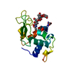
- PDB-1ljn: Crystal Structure of Turkey Egg Lysozyme Complex with Di-N-acetyl... -
+ Open data
- Basic information
| Entry | Database: PDB / ID: 1ljn | |||||||||
|---|---|---|---|---|---|---|---|---|---|---|
| Title | Crystal Structure of Turkey Egg Lysozyme Complex with Di-N-acetylchitobiose at 1.19A Resolution | |||||||||
 Components Components | lysozyme C | |||||||||
 Keywords Keywords | HYDROLASE / sugar complex / non-productive binding | |||||||||
| Function / homology |  Function and homology information Function and homology informationglycosaminoglycan binding / cell wall macromolecule catabolic process / lysozyme / lysozyme activity / defense response to Gram-negative bacterium / killing of cells of another organism / defense response to Gram-positive bacterium / extracellular space / identical protein binding / cytoplasm Similarity search - Function | |||||||||
| Biological species |  | |||||||||
| Method |  X-RAY DIFFRACTION / X-RAY DIFFRACTION /  MOLECULAR REPLACEMENT / Resolution: 1.19 Å MOLECULAR REPLACEMENT / Resolution: 1.19 Å | |||||||||
 Authors Authors | Harata, K. / Kanai, R. | |||||||||
 Citation Citation |  Journal: Proteins / Year: 2002 Journal: Proteins / Year: 2002Title: Crystallographic dissection of the thermal motion of protein-sugar complex. Authors: Harata, K. / Kanai, R. | |||||||||
| History |
|
- Structure visualization
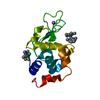
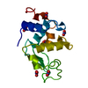
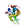
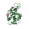
| Structure viewer | Molecule:  Molmil Molmil Jmol/JSmol Jmol/JSmol |
|---|
- Downloads & links
- Download
| PDBx/mmCIF format |  1ljn.cif.gz 1ljn.cif.gz | 74.5 KB | Display |  PDBx/mmCIF format PDBx/mmCIF format |
|---|---|---|---|---|
| PDB format |  pdb1ljn.ent.gz pdb1ljn.ent.gz | 55 KB | Display |  PDB format PDB format |
| PDBx/mmJSON format |  1ljn.json.gz 1ljn.json.gz | Tree view |  PDBx/mmJSON format PDBx/mmJSON format | |
| Others |  Other downloads Other downloads |
-Validation report
| Arichive directory |  https://data.pdbj.org/pub/pdb/validation_reports/lj/1ljn https://data.pdbj.org/pub/pdb/validation_reports/lj/1ljn ftp://data.pdbj.org/pub/pdb/validation_reports/lj/1ljn ftp://data.pdbj.org/pub/pdb/validation_reports/lj/1ljn | HTTPS FTP |
|---|
-Related structure data
| Related structure data | 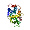 1lzyS S: Starting model for refinement |
|---|---|
| Similar structure data |
- Links
Links
- Assembly
| Deposited unit | 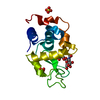
| ||||||||
|---|---|---|---|---|---|---|---|---|---|
| 1 |
| ||||||||
| Unit cell |
|
- Components
| #1: Protein | Mass: 14228.105 Da / Num. of mol.: 1 / Source method: isolated from a natural source / Source: (natural)  | ||||
|---|---|---|---|---|---|
| #2: Polysaccharide | 2-acetamido-2-deoxy-beta-D-glucopyranose-(1-4)-2-acetamido-2-deoxy-beta-D-glucopyranose Source method: isolated from a genetically manipulated source | ||||
| #3: Chemical | | #4: Water | ChemComp-HOH / | Has protein modification | Y | |
-Experimental details
-Experiment
| Experiment | Method:  X-RAY DIFFRACTION / Number of used crystals: 1 X-RAY DIFFRACTION / Number of used crystals: 1 |
|---|
- Sample preparation
| Crystal | Density Matthews: 1.94 Å3/Da / Density % sol: 36.63 % | ||||||||||||||||||||
|---|---|---|---|---|---|---|---|---|---|---|---|---|---|---|---|---|---|---|---|---|---|
| Crystal grow | Temperature: 293 K / Method: vapor diffusion / pH: 4.2 Details: ammonium sulfate, pH 4.2, VAPOR DIFFUSION, temperature 293K | ||||||||||||||||||||
| Crystal grow | *PLUS Method: batch method / Details: Harata, K., (1993) Acta Crystallogr., D49, 497. | ||||||||||||||||||||
| Components of the solutions | *PLUS
|
-Data collection
| Diffraction | Mean temperature: 286 K |
|---|---|
| Diffraction source | Source:  ROTATING ANODE / Type: ENRAF-NONIUS FR571 / Wavelength: 1.5418 Å ROTATING ANODE / Type: ENRAF-NONIUS FR571 / Wavelength: 1.5418 Å |
| Detector | Type: ENRAF-NONIUS FAST / Detector: AREA DETECTOR / Date: Nov 19, 1999 / Details: graphite monochromator |
| Radiation | Monochromator: graphite / Protocol: SINGLE WAVELENGTH / Monochromatic (M) / Laue (L): M / Scattering type: x-ray |
| Radiation wavelength | Wavelength: 1.5418 Å / Relative weight: 1 |
| Reflection | Resolution: 1.19→15.8 Å / Num. all: 35242 / Num. obs: 32310 / % possible obs: 91.7 % / Observed criterion σ(F): 0 / Observed criterion σ(I): 0 / Redundancy: 3.21 % / Rmerge(I) obs: 0.052 |
| Reflection shell | Resolution: 1.19→1.21 Å / Rmerge(I) obs: 0.206 / % possible all: 49.1 |
| Reflection | *PLUS Num. obs: 32432 / % possible obs: 92 % / Num. measured all: 103984 |
- Processing
| Software |
| |||||||||||||||||||||||||
|---|---|---|---|---|---|---|---|---|---|---|---|---|---|---|---|---|---|---|---|---|---|---|---|---|---|---|
| Refinement | Method to determine structure:  MOLECULAR REPLACEMENT MOLECULAR REPLACEMENTStarting model: PDB entry 1LZY Resolution: 1.19→15.8 Å / Isotropic thermal model: Anisotropic U / σ(F): 0 / Stereochemistry target values: Engh & Huber Details: For some water molecules, it can not be distinguished if the sites are occupied by one molecule or two, and thus the occupancies of the alternate conformations are greater than 1.00. For ...Details: For some water molecules, it can not be distinguished if the sites are occupied by one molecule or two, and thus the occupancies of the alternate conformations are greater than 1.00. For example, in HOH 238, occupancy(A)+occupancy(B)=1, occupancy(A)+occupancy(C)=1, and occupancy(B)=ocupancy(C).
| |||||||||||||||||||||||||
| Displacement parameters | Biso mean: 14 Å2 | |||||||||||||||||||||||||
| Refinement step | Cycle: LAST / Resolution: 1.19→15.8 Å
| |||||||||||||||||||||||||
| Refine LS restraints |
| |||||||||||||||||||||||||
| Software | *PLUS Name: SHELXL / Version: 97 / Classification: refinement | |||||||||||||||||||||||||
| Refinement | *PLUS % reflection Rfree: 5 % / Rfactor Rwork: 0.104 | |||||||||||||||||||||||||
| Solvent computation | *PLUS | |||||||||||||||||||||||||
| Displacement parameters | *PLUS |
 Movie
Movie Controller




 PDBj
PDBj


