 Movie
Movie Controller

[English] 日本語
 Yorodumi
- PDB-1jew: CRYO-EM STRUCTURE OF COXSACKIEVIRUS B3(M STRAIN) WITH ITS CELLULA... -
+ Open data

- Basic information
| Entry | Database: PDB / ID: 1jew | ||||||
|---|---|---|---|---|---|---|---|
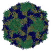
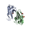
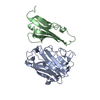
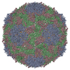
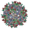
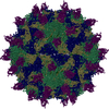
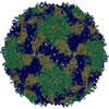
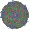
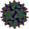
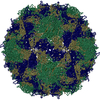
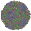
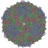
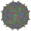
| Title | CRYO-EM STRUCTURE OF COXSACKIEVIRUS B3(M STRAIN) WITH ITS CELLULAR RECEPTOR, COXSACKIEVIRUS AND ADENOVIRUS RECEPTOR (CAR). | ||||||
 Components Components |
| ||||||
 Keywords Keywords | Virus/Receptor / COXSACKIEVIRUS B3 / CVB3 / CAR / CRYO-EM STRUCTURE / Icosahedral virus / Virus-Receptor COMPLEX | ||||||
| Function / homology |  Function and homology information Function and homology informationAV node cell-bundle of His cell adhesion involved in cell communication / cell adhesive protein binding involved in AV node cell-bundle of His cell communication / AV node cell to bundle of His cell communication / homotypic cell-cell adhesion / epithelial structure maintenance / regulation of AV node cell action potential / gamma-delta T cell activation / apicolateral plasma membrane / primordial germ cell migration / connexin binding ...AV node cell-bundle of His cell adhesion involved in cell communication / cell adhesive protein binding involved in AV node cell-bundle of His cell communication / AV node cell to bundle of His cell communication / homotypic cell-cell adhesion / epithelial structure maintenance / regulation of AV node cell action potential / gamma-delta T cell activation / apicolateral plasma membrane / primordial germ cell migration / connexin binding / cell-cell junction organization / transepithelial transport / heterophilic cell-cell adhesion / cardiac muscle cell development / symbiont-mediated suppression of host cytoplasmic pattern recognition receptor signaling pathway via inhibition of RIG-I activity / intercalated disc / bicellular tight junction / symbiont-mediated suppression of host cytoplasmic pattern recognition receptor signaling pathway via inhibition of MDA-5 activity / neutrophil chemotaxis / symbiont-mediated suppression of host cytoplasmic pattern recognition receptor signaling pathway via inhibition of MAVS activity / cell adhesion molecule binding / acrosomal vesicle / Cell surface interactions at the vascular wall / picornain 2A / filopodium / symbiont-mediated suppression of host mRNA export from nucleus / adherens junction / PDZ domain binding / neuromuscular junction / symbiont genome entry into host cell via pore formation in plasma membrane / picornain 3C / mitochondrion organization / T=pseudo3 icosahedral viral capsid / host cell cytoplasmic vesicle membrane / beta-catenin binding / integrin binding / Immunoregulatory interactions between a Lymphoid and a non-Lymphoid cell / cell-cell junction / cell junction / ribonucleoside triphosphate phosphatase activity / nucleoside-triphosphate phosphatase / heart development / virus receptor activity / growth cone / channel activity / actin cytoskeleton organization / cell body / monoatomic ion transmembrane transport / defense response to virus / basolateral plasma membrane / DNA replication / RNA helicase activity / neuron projection / endocytosis involved in viral entry into host cell / membrane raft / signaling receptor binding / symbiont-mediated activation of host autophagy / RNA-directed RNA polymerase / cysteine-type endopeptidase activity / viral RNA genome replication / RNA-directed RNA polymerase activity / DNA-templated transcription / virion attachment to host cell / host cell nucleus / structural molecule activity / protein-containing complex / proteolysis / : / RNA binding / extracellular region / zinc ion binding / nucleoplasm / ATP binding / identical protein binding / plasma membrane / cytoplasm Similarity search - Function | ||||||
| Biological species |  Homo sapiens (human) Homo sapiens (human)  Coxsackievirus B3 Coxsackievirus B3 | ||||||
| Method | ELECTRON MICROSCOPY / single particle reconstruction / cryo EM / Resolution: 22 Å | ||||||
 Authors Authors | Rossmann, M.G. / He, Y. | ||||||
 Citation Citation | Journal: Nat Struct Biol / Year: 2001 Title: Interaction of coxsackievirus B3 with the full length coxsackievirus-adenovirus receptor. Authors: Y He / P R Chipman / J Howitt / C M Bator / M A Whitt / T S Baker / R J Kuhn / C W Anderson / P Freimuth / M G Rossmann /  Abstract: Group B coxsackieviruses (CVB) utilize the coxsackievirus-adenovirus receptor (CAR) to recognize host cells. CAR is a membrane protein with two Ig-like extracellular domains (D1 and D2), a ...Group B coxsackieviruses (CVB) utilize the coxsackievirus-adenovirus receptor (CAR) to recognize host cells. CAR is a membrane protein with two Ig-like extracellular domains (D1 and D2), a transmembrane domain and a cytoplasmic domain. The three-dimensional structure of coxsackievirus B3 (CVB3) in complex with full length human CAR and also with the D1D2 fragment of CAR were determined to approximately 22 A resolution using cryo-electron microscopy (cryo-EM). Pairs of transmembrane domains of CAR associate with each other in a detergent cloud that mimics a cellular plasma membrane. This is the first view of a virus-receptor interaction at this resolution that includes the transmembrane and cytoplasmic portion of the receptor. CAR binds with the distal end of domain D1 in the canyon of CVB3, similar to how other receptor molecules bind to entero- and rhinoviruses. The previously described interface of CAR with the adenovirus knob protein utilizes a side surface of D1. #1: Journal: Science / Year: 1997Title: ISOLATION OF A COMMON RECEPTOR FOR COXSACKIE B VIRUSES AND ADENOVIRUSES 2 AND 5 Authors: Bergelson, J.M. / Cunningham, J.A. / Droguett, G. / Kurt-Jones, E.A. / Krithivas, A. / Hong, J.S. / Horwitz, M.S. / Crowell, R.L. / Finberg, R.W. #2: Journal: Science / Year: 1999Title: STRUCTURAL ANALYSIS OF THE MECHANISM OF ADENOVIRUS BINDING TO ITS HUMAN CELLULAR RECEPTOR CAR Authors: Bewley, M.C. / Springer, K. / Zhang, Y.B. / Freimuth, P. / Flanagan, J.M. #3: Journal: Structure / Year: 1995Title: THE STRUCTURE OF COXSACKIEVIRUS B3 AT 3.5A RESOLUTION Authors: Muckelbauer, J.K. / Kremer, M. / Minor, I. / Diana, G. / Dutko, F.J. / Groarke, J. / Pevear, D.C. / Rossman, M.G. | ||||||
| History |
| ||||||
| Remark 999 | SEQUENCE THE COXSACKIEVIRUS B3 USED IN THE EM EXPERIMENT IS M STRAIN, WHICH MIGHT BE DIFFERENT ...SEQUENCE THE COXSACKIEVIRUS B3 USED IN THE EM EXPERIMENT IS M STRAIN, WHICH MIGHT BE DIFFERENT SLIGHTLY WITH OTHER ISOLATES GIVING ARISE TO THE CONFLICTS. |
- Structure visualization
| Movie |
Movie viewer |
|---|---|
| Structure viewer | Molecule: MolmilJmol/JSmol |
- Downloads & links
-Download
| PDBx/mmCIF format | 1jew.cif.gz | 46.8 KB | Display | PDBx/mmCIF format |
|---|---|---|---|---|
| PDB format | pdb1jew.ent.gz | 24.4 KB | Display | PDB format |
| PDBx/mmJSON format | 1jew.json.gz | Tree view | PDBx/mmJSON format | |
| Others |  Other downloads Other downloads |
-Validation report
| Arichive directory | https://data.pdbj.org/pub/pdb/validation_reports/je/1jewftp://data.pdbj.org/pub/pdb/validation_reports/je/1jew | HTTPS FTP |
|---|
-Related structure data
| Related structure data | |
|---|---|
| Similar structure data |
-Links
 PDBj
PDBj


- Assembly
| Deposited unit | 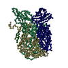
|
|---|---|
| 1 | x 60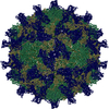
|
| 2 |
|
| 3 | x 5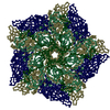
|
| 4 | x 6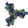
|
| 5 | 
|
| Symmetry | Point symmetry: (Hermann–Mauguin notation: 532 / Schoenflies symbol: I (icosahedral)) |
-Components
| #1: Protein | Mass: 13358.203 Da / Num. of mol.: 1 / Fragment: Residues 21-140 Source method: isolated from a genetically manipulated source Source: (gene. exp.) Homo sapiens (human) / Gene: CAR / Cell line (production host): A9 CELLS / Production host:  |
|---|---|
| #2: Protein | Mass: 31380.068 Da / Num. of mol.: 1 / Fragment: Residues 571-851 Source method: isolated from a genetically manipulated source Source: (gene. exp.) Coxsackievirus B3 (strain Woodruff) / Genus: Enterovirus / Species: Human enterovirus B / Strain: Woodruff / Description: COXSACKIEVIRUS B3 / Cell line (production host): HELA CELLS / Production host: Homo sapiens (human) / References: UniProt: Q66282 |
| #3: Protein | Mass: 28877.592 Da / Num. of mol.: 1 / Fragment: Residues 70-332 Source method: isolated from a genetically manipulated source Source: (gene. exp.) Coxsackievirus B3 (strain Woodruff) / Genus: Enterovirus / Species: Human enterovirus B / Strain: Woodruff / Description: COXSACKIEVIRUS B3 / Cell line (production host): HELA CELLS / Production host: Homo sapiens (human) / References: UniProt: Q66282 |
| #4: Protein | Mass: 26198.684 Da / Num. of mol.: 1 / Fragment: Residues 333-570 Source method: isolated from a genetically manipulated source Source: (gene. exp.) Coxsackievirus B3 (strain Woodruff) / Genus: Enterovirus / Species: Human enterovirus B / Strain: Woodruff / Description: COXSACKIEVIRUS B3 / Cell line (production host): HELA CELLS / Production host: Homo sapiens (human) / References: UniProt: Q66282 |
| #5: Protein | Mass: 7379.065 Da / Num. of mol.: 1 / Fragment: Residues 2-69 Source method: isolated from a genetically manipulated source Source: (gene. exp.) Coxsackievirus B3 (strain Woodruff) / Genus: Enterovirus / Species: Human enterovirus B / Strain: Woodruff / Description: COXSACKIEVIRUS B3 / Cell line (production host): HELA CELLS / Production host: Homo sapiens (human) / References: UniProt: Q66282 |
-Experimental details
-Experiment
| Experiment | Method: ELECTRON MICROSCOPY |
|---|---|
| EM experiment | Aggregation state: PARTICLE / 3D reconstruction method: single particle reconstruction |
- Sample preparation
| Component | Name: COXSACKIEVIRUS B3(M STRAIN) WITH ITS CELLULAR RECEPTOR Type: VIRUS | ||||||||||||
|---|---|---|---|---|---|---|---|---|---|---|---|---|---|
| Source (natural) |
| ||||||||||||
| Source (recombinant) |
| ||||||||||||
| Buffer solution | pH: 7.5 | ||||||||||||
| Specimen | Embedding applied: NO / Shadowing applied: NO / Staining applied: NO / Vitrification applied: YES | ||||||||||||
| Vitrification | Details: CVB3 WAS INCUBATED WITH CAR SAMPLE FOR 1 HOURS AT 25 DEGREES CELSIUS (298 KELVIN) USING A FOUR-FOLD EXCESS OF CAR FOR EACH OF THE SIXTY POSSIBLE BINDING SITES PER VIRION. AFTER INCUBATION, ...Details: CVB3 WAS INCUBATED WITH CAR SAMPLE FOR 1 HOURS AT 25 DEGREES CELSIUS (298 KELVIN) USING A FOUR-FOLD EXCESS OF CAR FOR EACH OF THE SIXTY POSSIBLE BINDING SITES PER VIRION. AFTER INCUBATION, SAMPLES WERE PREPARED AS THIN LAYERS OF VITREOUS ICE AND MAINTAINED AT NEAR LIQUID NITROGEN TEMPERATURE IN THE ELECTRON MICROSCOPE WITH A GATAN 626 CRYOTRANSFER HOLDER. | ||||||||||||
| Crystal grow | Temperature: 298 K / pH: 7.5 Details: WARNING: THIS IS AN CRYO-ELECTRON MICROSCOPY MODEL DEPOSITION. CRYO-EM INFORMATION HAS BEEN INCLUDED IN THE PDB FILE., pH 7.5, ELECTRON MICROSCOPY RECONSTRUCTION, temperature 298K | ||||||||||||
| Crystal grow | *PLUS Method: electron microscopy |
- Electron microscopy imaging
| Microscopy | Model: FEI/PHILIPS CM300FEG/T / Date: Jun 1, 2000 |
|---|---|
| Electron gun | Electron source:  FIELD EMISSION GUN / Accelerating voltage: 300 kV / Illumination mode: FLOOD BEAM FIELD EMISSION GUN / Accelerating voltage: 300 kV / Illumination mode: FLOOD BEAM |
| Electron lens | Mode: BRIGHT FIELD / Nominal magnification: 61000 X / Nominal defocus max: 4600 nm / Nominal defocus min: 1500 nm |
| Specimen holder | Temperature: 120 K |
| Image recording | Electron dose: 20 e/Å2 / Film or detector model: KODAK SO-163 FILM |
- Processing
| EM software | Name: PURDUE PROGRAMS / Category: 3D reconstruction | ||||||||||||
|---|---|---|---|---|---|---|---|---|---|---|---|---|---|
| Symmetry | Point symmetry: I (icosahedral) | ||||||||||||
| 3D reconstruction | Method: COMMON-LINES AND POLAR-FOURIER-TRANSFORM FULLER ET AL. 1996, J.STRUC.BIOL.c 116, 48-55; BAKER AND CHENG, 1996, J.STRUC.BIOL. 116, 120-130 Resolution: 22 Å / Resolution method: OTHER / Num. of particles: 635 / Actual pixel size: 2.3 Å Magnification calibration: THE PIXEL SIZE OF THE CRYO-EM MAP WAS CALIBRATED AGAINST A LOW RESOLUTION DENSITY MAP CALCULATED FROM THE CRYSTAL STRUCTURE OF COXSACKIEVIRUS B3. DENSITIES WERE COMPARED BY ...Magnification calibration: THE PIXEL SIZE OF THE CRYO-EM MAP WAS CALIBRATED AGAINST A LOW RESOLUTION DENSITY MAP CALCULATED FROM THE CRYSTAL STRUCTURE OF COXSACKIEVIRUS B3. DENSITIES WERE COMPARED BY CROSS-CORRELATION WITHIN A SPHERICAL SHELL OF INTERNAL RADIUS CORRELATION WITHIN A SPHERICAL SHELL OF INTERNAL RADIUS 110 ANGSTROMS AND EXTERNAL RADIUS OF 145 ANGSTROMS. Details: THE RESOLUTION OF THE FINAL RECONSTRUCTED DENSITY WAS DETERMINED TO BE AT LEAST 22 ANGSTROMS, AS MEASURED BY RANDOMLY SPLITTING THE PARTICLES INTO TWO SETS AND COMPARING STRUCTURE FACTORS ...Details: THE RESOLUTION OF THE FINAL RECONSTRUCTED DENSITY WAS DETERMINED TO BE AT LEAST 22 ANGSTROMS, AS MEASURED BY RANDOMLY SPLITTING THE PARTICLES INTO TWO SETS AND COMPARING STRUCTURE FACTORS OBTAINED FROM SEPARATE RECONSTRUCTIONS (BAKER ET AL. 1991, BIOPHYS.J. 60, 1445-1456). THE EIGENVALUE SPECTRUM GAVE AN INDICATION OF THE RANDOMNESS OF THE DATA THAT WAS INCLUDED IN THE RECONSTRUCTION. THE COMPLETENESS OF THE DATA WAS VERIFIED IN THAT ALL EIGENVALUES EXCEEDED 1.0. Symmetry type: POINT | ||||||||||||
| Atomic model building | Protocol: OTHER / Space: REAL Details: METHOD--Acta Cryst. D56, 1341 REFINEMENT PROTOCOL--EMFIT DETAILS--THE DIFFERENCE MAP BETWEEN CVB3-CAR COMPLEX AND NATIVE CVB WAS CALCULATED AND THEN FITTED WITH THE N-TERMINAL DOMAIN OF CAR ...Details: METHOD--Acta Cryst. D56, 1341 REFINEMENT PROTOCOL--EMFIT DETAILS--THE DIFFERENCE MAP BETWEEN CVB3-CAR COMPLEX AND NATIVE CVB WAS CALCULATED AND THEN FITTED WITH THE N-TERMINAL DOMAIN OF CAR (1KAC, B-CHAIN). THE AUTOMATIC FITTING WAS DONE USI PROGRAM EMFIT DESCRIBED IN ACTA CRYST. D56, 1341-1349. THE CRYSTAL STRUCTURE OF POLIOVIRUS WAS PLACED INTO THE | ||||||||||||
| Refinement step | Cycle: LAST
|
