 Movie
Movie Controller

[English] 日本語
 Yorodumi
- PDB-1kac: KNOB DOMAIN FROM ADENOVIRUS SEROTYPE 12 IN COMPLEX WITH DOMAIN 1 ... -
+ Open data

- Basic information
| Entry | Database: PDB / ID: 1kac | ||||||
|---|---|---|---|---|---|---|---|
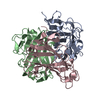
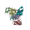
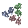
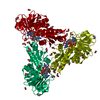
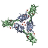
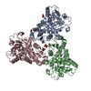
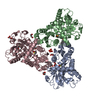
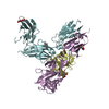
| Title | KNOB DOMAIN FROM ADENOVIRUS SEROTYPE 12 IN COMPLEX WITH DOMAIN 1 OF ITS CELLULAR RECEPTOR CAR | ||||||
 Components Components |
| ||||||
 Keywords Keywords | Viral protein/receptor / ADHESION PROTEIN RECEPTOR COMPLEX / VIRAL PROTEIN-RECEPTOR COMPLEX | ||||||
| Function / homology |  Function and homology information Function and homology informationAV node cell-bundle of His cell adhesion involved in cell communication / cell adhesive protein binding involved in AV node cell-bundle of His cell communication / homotypic cell-cell adhesion / AV node cell to bundle of His cell communication / epithelial structure maintenance / regulation of AV node cell action potential / primordial germ cell migration / apicolateral plasma membrane / gamma-delta T cell activation / cell-cell junction organization ...AV node cell-bundle of His cell adhesion involved in cell communication / cell adhesive protein binding involved in AV node cell-bundle of His cell communication / homotypic cell-cell adhesion / AV node cell to bundle of His cell communication / epithelial structure maintenance / regulation of AV node cell action potential / primordial germ cell migration / apicolateral plasma membrane / gamma-delta T cell activation / cell-cell junction organization / connexin binding / cardiac muscle cell development / transepithelial transport / adhesion receptor-mediated virion attachment to host cell / heterophilic cell-cell adhesion / intercalated disc / bicellular tight junction / neutrophil chemotaxis / cell adhesion molecule binding / acrosomal vesicle / Cell surface interactions at the vascular wall / mitochondrion organization / filopodium / adherens junction / PDZ domain binding / neuromuscular junction / cell junction / beta-catenin binding / integrin binding / Immunoregulatory interactions between a Lymphoid and a non-Lymphoid cell / heart development / cell-cell junction / viral capsid / growth cone / virus receptor activity / actin cytoskeleton organization / cell body / defense response to virus / basolateral plasma membrane / cell adhesion / neuron projection / membrane raft / signaling receptor binding / symbiont entry into host cell / host cell nucleus / protein-containing complex / : / extracellular region / identical protein binding / plasma membrane / cytoplasm Similarity search - Function | ||||||
| Biological species |  Human adenovirus 12 Human adenovirus 12 Homo sapiens (human) Homo sapiens (human) | ||||||
| Method |  X-RAY DIFFRACTION / SYNCHROTRON / OTHER / Resolution: 2.6 Å X-RAY DIFFRACTION / SYNCHROTRON / OTHER / Resolution: 2.6 Å | ||||||
 Authors Authors | Bewley, M.C. / Springer, K. / Zhang, Y.B. / Freimuth, P. / Flanagan, J.M. | ||||||
 Citation Citation | Journal: Science / Year: 1999 Title: Structural analysis of the mechanism of adenovirus binding to its human cellular receptor, CAR. Authors: Bewley, M.C. / Springer, K. / Zhang, Y.B. / Freimuth, P. / Flanagan, J.M. | ||||||
| History |
|
- Structure visualization
| Structure viewer | Molecule: MolmilJmol/JSmol |
|---|
- Downloads & links
-Download
| PDBx/mmCIF format | 1kac.cif.gz | 73.1 KB | Display | PDBx/mmCIF format |
|---|---|---|---|---|
| PDB format | pdb1kac.ent.gz | 54.6 KB | Display | PDB format |
| PDBx/mmJSON format | 1kac.json.gz | Tree view | PDBx/mmJSON format | |
| Others |  Other downloads Other downloads |
-Validation report
| Arichive directory | https://data.pdbj.org/pub/pdb/validation_reports/ka/1kacftp://data.pdbj.org/pub/pdb/validation_reports/ka/1kac | HTTPS FTP |
|---|
-Related structure data
-Links
 PDBj
PDBj


- Assembly
| Deposited unit | 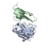
| ||||||||
|---|---|---|---|---|---|---|---|---|---|
| 1 | 
| ||||||||
| Unit cell |
|
-Components
| #1: Protein | Mass: 19944.477 Da / Num. of mol.: 1 / Fragment: KNOB Source method: isolated from a genetically manipulated source Source: (gene. exp.) Human adenovirus 12 / Genus: Mastadenovirus / Species: Human adenovirus A / Variant: SEROTYPE 12 / Cell line (production host): BL21(DE3) / Production host:  |
|---|---|
| #2: Protein | Mass: 13640.500 Da / Num. of mol.: 1 / Fragment: DOMAIN 1 Source method: isolated from a genetically manipulated source Source: (gene. exp.) Homo sapiens (human) / References: UniProt: P78310 |
| #3: Water | ChemComp-HOH /  Mass: 18.015 Da / Num. of mol.: 70 / Source method: isolated from a natural source / Formula: H2O Mass: 18.015 Da / Num. of mol.: 70 / Source method: isolated from a natural source / Formula: H2O |
| Has protein modification | Y |
-Experimental details
-Experiment
| Experiment | Method: X-RAY DIFFRACTION / Number of used crystals: 1 |
|---|
- Sample preparation
| Crystal | Density Matthews: 5.6 Å3/Da / Density % sol: 78 % | ||||||||||||||||||
|---|---|---|---|---|---|---|---|---|---|---|---|---|---|---|---|---|---|---|---|
| Crystal grow | pH: 6.2 / Details: 900MM AMMONIUM SULPHATE ON 100MM MES, PH 6.2 | ||||||||||||||||||
| Crystal | *PLUS | ||||||||||||||||||
| Crystal grow | *PLUS Method: vapor diffusion, sitting drop | ||||||||||||||||||
| Components of the solutions | *PLUS
|
-Data collection
| Diffraction | Mean temperature: 99 K |
|---|---|
| Diffraction source | Source: SYNCHROTRON / Site: NSLS  / Beamline: X25 / Wavelength: 1 / Beamline: X25 / Wavelength: 1 |
| Radiation | Protocol: SINGLE WAVELENGTH / Monochromatic (M) / Laue (L): M / Scattering type: x-ray |
| Radiation wavelength | Wavelength: 1 Å / Relative weight: 1 |
| Reflection | Resolution: 2.6→40 Å / Num. obs: 24636 / % possible obs: 100 % / Redundancy: 14.2 % / Rmerge(I) obs: 0.7 |
| Reflection shell | Resolution: 2.6→2.7 Å / Rmerge(I) obs: 0.35 / % possible all: 100 |
| Reflection shell | *PLUS % possible obs: 100 % / Mean I/σ(I) obs: 6 |
- Processing
| Software |
| ||||||||||||||||||||||||||||||||||||||||||||||||||||||||||||
|---|---|---|---|---|---|---|---|---|---|---|---|---|---|---|---|---|---|---|---|---|---|---|---|---|---|---|---|---|---|---|---|---|---|---|---|---|---|---|---|---|---|---|---|---|---|---|---|---|---|---|---|---|---|---|---|---|---|---|---|---|---|
| Refinement | Method to determine structure: OTHER / Resolution: 2.6→20 Å / Isotropic thermal model: RESTRAINED / Cross valid method: THROUGHOUT / σ(F): 0
| ||||||||||||||||||||||||||||||||||||||||||||||||||||||||||||
| Solvent computation | Bsol: 61 Å2 / ksol: 0.43 e/Å3 | ||||||||||||||||||||||||||||||||||||||||||||||||||||||||||||
| Displacement parameters | Biso mean: 44.9 Å2
| ||||||||||||||||||||||||||||||||||||||||||||||||||||||||||||
| Refinement step | Cycle: LAST / Resolution: 2.6→20 Å
| ||||||||||||||||||||||||||||||||||||||||||||||||||||||||||||
| Refine LS restraints |
| ||||||||||||||||||||||||||||||||||||||||||||||||||||||||||||
| Xplor file | Serial no: 1 / Param file: PROTEIN_REP.PARAM | ||||||||||||||||||||||||||||||||||||||||||||||||||||||||||||
| Software | *PLUS Name: CNS / Version: 5 / Classification: refinement | ||||||||||||||||||||||||||||||||||||||||||||||||||||||||||||
| Refinement | *PLUS Highest resolution: 2.6 Å / Lowest resolution: 20 Å / σ(F): 0 / % reflection Rfree: 2.9 % / Rfactor obs: 0.22 / Rfactor Rfree: 0.25 / Rfactor Rwork: 0.22 | ||||||||||||||||||||||||||||||||||||||||||||||||||||||||||||
| Solvent computation | *PLUS | ||||||||||||||||||||||||||||||||||||||||||||||||||||||||||||
| Displacement parameters | *PLUS Biso mean: 44.9 Å2 | ||||||||||||||||||||||||||||||||||||||||||||||||||||||||||||
| Refine LS restraints | *PLUS Type: c_angle_deg / Dev ideal: 1.7 |
