 Movie
Movie Controller

[English] 日本語
 Yorodumi
- PDB-6tm3: Structure of methylene-tetrahydromethanopterin dehydrogenase from... -
+ Open data

- Basic information
| Entry | Database: PDB / ID: 6tm3 | ||||||||||||
|---|---|---|---|---|---|---|---|---|---|---|---|---|---|
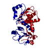
| Title | Structure of methylene-tetrahydromethanopterin dehydrogenase from Methylorubrum extorquens AM1 in a close conformation containing NADP+ and methylene-H4MPT | ||||||||||||
 Components Components | Bifunctional protein MdtA | ||||||||||||
 Keywords Keywords | OXIDOREDUCTASE / One-carbon metabolism / Enzyme catalysis / Dehydrogenase / Conformational changes / Methylotrophy | ||||||||||||
| Function / homology |  Function and homology information Function and homology informationOxidoreductases; Acting on the CH-NH group of donors; With NAD+ or NADP+ as acceptor / methylenetetrahydrofolate dehydrogenase (NADP+) / methylenetetrahydrofolate dehydrogenase (NADP+) activity / formaldehyde catabolic process / one-carbon metabolic process / cytoplasm Similarity search - Function | ||||||||||||
| Biological species |  Methylorubrum extorquens AM1 (bacteria) Methylorubrum extorquens AM1 (bacteria) | ||||||||||||
| Method |  X-RAY DIFFRACTION / SYNCHROTRON / MOLECULAR REPLACEMENT / Resolution: 1.08 Å X-RAY DIFFRACTION / SYNCHROTRON / MOLECULAR REPLACEMENT / Resolution: 1.08 Å | ||||||||||||
 Authors Authors | Wagner, T. / Huang, G. / Demmer, U. / Warkentin, E. / Ermler, U. / Shima, S. | ||||||||||||
| Funding support |  Germany, Germany,  China, 3items China, 3items
| ||||||||||||
 Citation Citation | Journal: J.Mol.Biol. / Year: 2020 Title: The Hydride Transfer Process in NADP-dependent Methylene-tetrahydromethanopterin Dehydrogenase. Authors: Huang, G. / Wagner, T. / Demmer, U. / Warkentin, E. / Ermler, U. / Shima, S. | ||||||||||||
| History |
|
- Structure visualization
| Structure viewer | Molecule: MolmilJmol/JSmol |
|---|
- Downloads & links
-Download
| PDBx/mmCIF format | 6tm3.cif.gz | 161.9 KB | Display | PDBx/mmCIF format |
|---|---|---|---|---|
| PDB format | pdb6tm3.ent.gz | 124.7 KB | Display | PDB format |
| PDBx/mmJSON format | 6tm3.json.gz | Tree view | PDBx/mmJSON format | |
| Others |  Other downloads Other downloads |
-Validation report
| Arichive directory | https://data.pdbj.org/pub/pdb/validation_reports/tm/6tm3ftp://data.pdbj.org/pub/pdb/validation_reports/tm/6tm3 | HTTPS FTP |
|---|
-Related structure data
| Related structure data | 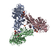 6tgeC  6tlkC  6yk9C 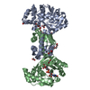 6ykaC 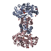 6ykbC 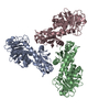 1lu9S S: Starting model for refinement C: citing same article ( |
|---|---|
| Similar structure data |

-Links
 PDBj
PDBj
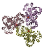
- Assembly
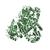
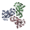
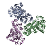
| Deposited unit | 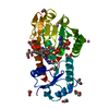
| ||||||||||||||||||||||||||||||
|---|---|---|---|---|---|---|---|---|---|---|---|---|---|---|---|---|---|---|---|---|---|---|---|---|---|---|---|---|---|---|---|
| 1 | 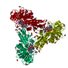
| ||||||||||||||||||||||||||||||
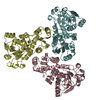
| Unit cell |
| ||||||||||||||||||||||||||||||
| Components on special symmetry positions |
|
-Components
-Protein , 1 types, 1 molecules A
| #1: Protein | Mass: 31512.734 Da / Num. of mol.: 1 Source method: isolated from a genetically manipulated source Source: (gene. exp.) Methylorubrum extorquens AM1 (bacteria)Gene: mtdA, MexAM1_META1p1728 / Plasmid: pET-24b(+) Details (production host): The synthesized DNA fragment was inserted into the expression vector pET-24b (+) at the NdeI and SalI restriction-enzyme digestion-sites. Production host: References: UniProt: P55818, Oxidoreductases; Acting on the CH-NH group of donors; With NAD+ or NADP+ as acceptor, methylenetetrahydrofolate dehydrogenase (NADP+) |
|---|
-Non-polymers , 6 types, 520 molecules 

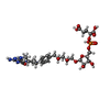


| #2: Chemical | ChemComp-NAP /  Mass: 743.405 Da / Num. of mol.: 1 / Source method: obtained synthetically / Formula: C21H28N7O17P3 / Feature type: SUBJECT OF INVESTIGATION Mass: 743.405 Da / Num. of mol.: 1 / Source method: obtained synthetically / Formula: C21H28N7O17P3 / Feature type: SUBJECT OF INVESTIGATION | ||||
|---|---|---|---|---|---|
| #3: Chemical | ChemComp-GOL /  Mass: 92.094 Da / Num. of mol.: 1 / Source method: obtained synthetically / Formula: C3H8O3 Mass: 92.094 Da / Num. of mol.: 1 / Source method: obtained synthetically / Formula: C3H8O3 | ||||
| #4: Chemical | ChemComp-H4M /  Mass: 788.693 Da / Num. of mol.: 1 / Source method: obtained synthetically / Formula: C31H45N6O16P / Feature type: SUBJECT OF INVESTIGATION Mass: 788.693 Da / Num. of mol.: 1 / Source method: obtained synthetically / Formula: C31H45N6O16P / Feature type: SUBJECT OF INVESTIGATION | ||||
| #5: Chemical | ChemComp-EDO /  Mass: 62.068 Da / Num. of mol.: 14 / Source method: obtained synthetically / Formula: C2H6O2 Mass: 62.068 Da / Num. of mol.: 14 / Source method: obtained synthetically / Formula: C2H6O2#6: Chemical |  Mass: 6.941 Da / Num. of mol.: 2 / Source method: obtained synthetically / Formula: Li Mass: 6.941 Da / Num. of mol.: 2 / Source method: obtained synthetically / Formula: Li#7: Water | ChemComp-HOH / | Mass: 18.015 Da / Num. of mol.: 501 / Source method: isolated from a natural source / Formula: H2O |
-Details
| Has ligand of interest | Y |
|---|
-Experimental details
-Experiment
| Experiment | Method: X-RAY DIFFRACTION / Number of used crystals: 1 |
|---|
- Sample preparation
| Crystal | Density Matthews: 2.63 Å3/Da / Density % sol: 53.22 % |
|---|---|
| Crystal grow | Temperature: 281.15 K / Method: vapor diffusion, sitting drop / pH: 8 Details: The crystallized sample was 25 mg/ml MtdA supplemented with 2.5 mM methenyl-H4MPT, 2 mM NADPH in 25 mM Tris/HCl pH 7.5, 150 mM NaCl, 5% glycerol and 2 mM dithiothreitol. 0.7 ul protein ...Details: The crystallized sample was 25 mg/ml MtdA supplemented with 2.5 mM methenyl-H4MPT, 2 mM NADPH in 25 mM Tris/HCl pH 7.5, 150 mM NaCl, 5% glycerol and 2 mM dithiothreitol. 0.7 ul protein sample was mixed with 0.7 ul of the reservoir solution containing 30% w/v polyethylene glycol monomethyl ether 5000, 100 mM Tris pH 8.0 and 200 mM lithium sulfate. PH range: / / Temp details: / |
-Data collection
| Diffraction | Mean temperature: 100 K / Serial crystal experiment: N | |||||||||||||||||||||||||||||||||||||||||||||||||||||||||||||||||||||||||||||||||||||||||||||||||||||||||||||||||||||||||
|---|---|---|---|---|---|---|---|---|---|---|---|---|---|---|---|---|---|---|---|---|---|---|---|---|---|---|---|---|---|---|---|---|---|---|---|---|---|---|---|---|---|---|---|---|---|---|---|---|---|---|---|---|---|---|---|---|---|---|---|---|---|---|---|---|---|---|---|---|---|---|---|---|---|---|---|---|---|---|---|---|---|---|---|---|---|---|---|---|---|---|---|---|---|---|---|---|---|---|---|---|---|---|---|---|---|---|---|---|---|---|---|---|---|---|---|---|---|---|---|---|---|---|
| Diffraction source | Source: SYNCHROTRON / Site: SLS  / Beamline: X10SA / Wavelength: 1 Å / Beamline: X10SA / Wavelength: 1 Å | |||||||||||||||||||||||||||||||||||||||||||||||||||||||||||||||||||||||||||||||||||||||||||||||||||||||||||||||||||||||||
| Detector | Type: DECTRIS PILATUS 6M-F / Detector: PIXEL / Date: Feb 4, 2018 | |||||||||||||||||||||||||||||||||||||||||||||||||||||||||||||||||||||||||||||||||||||||||||||||||||||||||||||||||||||||||
| Radiation | Protocol: SINGLE WAVELENGTH / Monochromatic (M) / Laue (L): M / Scattering type: x-ray | |||||||||||||||||||||||||||||||||||||||||||||||||||||||||||||||||||||||||||||||||||||||||||||||||||||||||||||||||||||||||
| Radiation wavelength | Wavelength: 1 Å / Relative weight: 1 | |||||||||||||||||||||||||||||||||||||||||||||||||||||||||||||||||||||||||||||||||||||||||||||||||||||||||||||||||||||||||
| Reflection | Resolution: 1.08→88.922 Å / Num. all: 143688 / Num. obs: 143688 / % possible obs: 99.6 % / Redundancy: 10 % / Rpim(I) all: 0.016 / Rrim(I) all: 0.051 / Rsym value: 0.049 / Net I/av σ(I): 10.3 / Net I/σ(I): 25.1 / Num. measured all: 1439110 | |||||||||||||||||||||||||||||||||||||||||||||||||||||||||||||||||||||||||||||||||||||||||||||||||||||||||||||||||||||||||
| Reflection shell | Diffraction-ID: 1
|
- Processing
| Software |
| ||||||||||||||||||||||||||||||||||||||||||||||||||||||||||||||||||||||||||||||||||||||||||||||||||||||||||||||||||||||||||||||||||||||||||||||||||||||||||||||||||||||||||||||||||||||||||
|---|---|---|---|---|---|---|---|---|---|---|---|---|---|---|---|---|---|---|---|---|---|---|---|---|---|---|---|---|---|---|---|---|---|---|---|---|---|---|---|---|---|---|---|---|---|---|---|---|---|---|---|---|---|---|---|---|---|---|---|---|---|---|---|---|---|---|---|---|---|---|---|---|---|---|---|---|---|---|---|---|---|---|---|---|---|---|---|---|---|---|---|---|---|---|---|---|---|---|---|---|---|---|---|---|---|---|---|---|---|---|---|---|---|---|---|---|---|---|---|---|---|---|---|---|---|---|---|---|---|---|---|---|---|---|---|---|---|---|---|---|---|---|---|---|---|---|---|---|---|---|---|---|---|---|---|---|---|---|---|---|---|---|---|---|---|---|---|---|---|---|---|---|---|---|---|---|---|---|---|---|---|---|---|---|---|---|---|
| Refinement | Method to determine structure: MOLECULAR REPLACEMENT Starting model: 1LU9 Resolution: 1.08→41.918 Å / SU ML: 0.11 / Cross valid method: THROUGHOUT / σ(F): 1.33 / Phase error: 13.76 / Stereochemistry target values: ML
| ||||||||||||||||||||||||||||||||||||||||||||||||||||||||||||||||||||||||||||||||||||||||||||||||||||||||||||||||||||||||||||||||||||||||||||||||||||||||||||||||||||||||||||||||||||||||||
| Solvent computation | Shrinkage radii: 0.9 Å / VDW probe radii: 1.11 Å / Solvent model: FLAT BULK SOLVENT MODEL | ||||||||||||||||||||||||||||||||||||||||||||||||||||||||||||||||||||||||||||||||||||||||||||||||||||||||||||||||||||||||||||||||||||||||||||||||||||||||||||||||||||||||||||||||||||||||||
| Displacement parameters | Biso max: 66.59 Å2 / Biso mean: 18.0707 Å2 / Biso min: 9.27 Å2 | ||||||||||||||||||||||||||||||||||||||||||||||||||||||||||||||||||||||||||||||||||||||||||||||||||||||||||||||||||||||||||||||||||||||||||||||||||||||||||||||||||||||||||||||||||||||||||
| Refinement step | Cycle: final / Resolution: 1.08→41.918 Å
| ||||||||||||||||||||||||||||||||||||||||||||||||||||||||||||||||||||||||||||||||||||||||||||||||||||||||||||||||||||||||||||||||||||||||||||||||||||||||||||||||||||||||||||||||||||||||||
| LS refinement shell | Refine-ID: X-RAY DIFFRACTION / Rfactor Rfree error: 0
|
