 Movie
Movie Controller


+ Open data

- Basic information
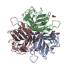
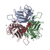
| Entry | Database: PDB / ID: 1nob | ||||||
|---|---|---|---|---|---|---|---|
| Title | KNOB DOMAIN FROM ADENOVIRUS SEROTYPE 12 | ||||||
 Components Components | PROTEIN (FIBER KNOB PROTEIN) | ||||||
 Keywords Keywords | VIRAL PROTEIN / RECEPTOR BINDING | ||||||
| Function / homology |  Function and homology information Function and homology informationadhesion receptor-mediated virion attachment to host cell / viral capsid / cell adhesion / symbiont entry into host cell / host cell nucleus Similarity search - Function | ||||||
| Biological species |  Human adenovirus 12 Human adenovirus 12 | ||||||
| Method |  X-RAY DIFFRACTION / SYNCHROTRON / MOLECULAR REPLACEMENT / Resolution: 2.6 Å X-RAY DIFFRACTION / SYNCHROTRON / MOLECULAR REPLACEMENT / Resolution: 2.6 Å | ||||||
 Authors Authors | Bewley, M.C. / Springer, K. / Zhang, Y.B. / Freimuth, P. / Flanagan, J.M. | ||||||
 Citation Citation | Journal: Science / Year: 1999 Title: Structural analysis of the mechanism of adenovirus binding to its human cellular receptor, CAR. Authors: Bewley, M.C. / Springer, K. / Zhang, Y.B. / Freimuth, P. / Flanagan, J.M. | ||||||
| History |
|
- Structure visualization
| Structure viewer | Molecule: MolmilJmol/JSmol |
|---|
- Downloads & links
-Download
| PDBx/mmCIF format | 1nob.cif.gz | 209.8 KB | Display | PDBx/mmCIF format |
|---|---|---|---|---|
| PDB format | pdb1nob.ent.gz | 170.9 KB | Display | PDB format |
| PDBx/mmJSON format | 1nob.json.gz | Tree view | PDBx/mmJSON format | |
| Others |  Other downloads Other downloads |
-Validation report
| Arichive directory | https://data.pdbj.org/pub/pdb/validation_reports/no/1nobftp://data.pdbj.org/pub/pdb/validation_reports/no/1nob | HTTPS FTP |
|---|
-Related structure data
| Related structure data | 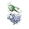 1kacC  1knbS C: citing same article ( S: Starting model for refinement |
|---|---|
| Similar structure data |


-Links
 PDBj
PDBj
- Assembly
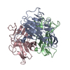
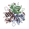
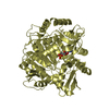
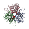
| Deposited unit | 
| ||||||||||||||||||||||||
|---|---|---|---|---|---|---|---|---|---|---|---|---|---|---|---|---|---|---|---|---|---|---|---|---|---|
| 1 | 
| ||||||||||||||||||||||||
| 2 | 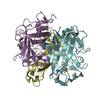
| ||||||||||||||||||||||||
| Unit cell |
| ||||||||||||||||||||||||
| Noncrystallographic symmetry (NCS) | NCS oper:
|
-Components
| #1: Protein | Mass: 19944.477 Da / Num. of mol.: 6 / Fragment: KNOB Source method: isolated from a genetically manipulated source Source: (gene. exp.) Human adenovirus 12 / Genus: Mastadenovirus / Species: Human adenovirus A / Variant: SEROTYPE 12 / Cell line (production host): BL21(DE3) / Production host:  |
|---|
-Experimental details
-Experiment
| Experiment | Method: X-RAY DIFFRACTION / Number of used crystals: 1 |
|---|
- Sample preparation
| Crystal | Density Matthews: 2 Å3/Da / Density % sol: 40 % |
|---|---|
| Crystal grow | pH: 7 / Details: 26% PEG 3350, pH 7.0 |
| Crystal | *PLUS |
| Crystal grow | *PLUS Method: vapor diffusion, sitting drop |
| Components of the solutions | *PLUS Conc.: 26 % / Common name: PEG3350 |
-Data collection
| Diffraction | Mean temperature: 99 K |
|---|---|
| Diffraction source | Source: SYNCHROTRON / Site: NSLS  / Beamline: X25 / Wavelength: 1 / Beamline: X25 / Wavelength: 1 |
| Detector | Type: BRANDEIS / Detector: CCD |
| Radiation | Protocol: SINGLE WAVELENGTH / Monochromatic (M) / Laue (L): M / Scattering type: x-ray |
| Radiation wavelength | Wavelength: 1 Å / Relative weight: 1 |
| Reflection | Resolution: 2.6→30 Å / Num. obs: 27987 / % possible obs: 100 % / Redundancy: 2.5 % / Rmerge(I) obs: 0.1 / Net I/σ(I): 10 |
| Reflection shell | Resolution: 2.6→2.7 Å / Rmerge(I) obs: 0.22 / % possible all: 100 |
| Reflection | *PLUS Rmerge(I) obs: 0.1 |
| Reflection shell | *PLUS % possible obs: 100 % / Mean I/σ(I) obs: 3 |
- Processing
| Software |
| ||||||||||||||||||||||||||||||||||||||||||||||||||||||||||||
|---|---|---|---|---|---|---|---|---|---|---|---|---|---|---|---|---|---|---|---|---|---|---|---|---|---|---|---|---|---|---|---|---|---|---|---|---|---|---|---|---|---|---|---|---|---|---|---|---|---|---|---|---|---|---|---|---|---|---|---|---|---|
| Refinement | Method to determine structure: MOLECULAR REPLACEMENT Starting model: 1KNB Resolution: 2.6→20 Å / Isotropic thermal model: RESTRAINED / Cross valid method: THROUGHOUT / σ(F): 0
| ||||||||||||||||||||||||||||||||||||||||||||||||||||||||||||
| Solvent computation | Bsol: 63 Å2 / ksol: 0.43 e/Å3 | ||||||||||||||||||||||||||||||||||||||||||||||||||||||||||||
| Displacement parameters | Biso mean: 18.02 Å2
| ||||||||||||||||||||||||||||||||||||||||||||||||||||||||||||
| Refinement step | Cycle: LAST / Resolution: 2.6→20 Å
| ||||||||||||||||||||||||||||||||||||||||||||||||||||||||||||
| Refine LS restraints |
| ||||||||||||||||||||||||||||||||||||||||||||||||||||||||||||
| Refine LS restraints NCS | NCS model details: RESTRAINTS | ||||||||||||||||||||||||||||||||||||||||||||||||||||||||||||
| Xplor file | Serial no: 1 / Param file: PROTEIN_REP.PARAM | ||||||||||||||||||||||||||||||||||||||||||||||||||||||||||||
| Software | *PLUS Name: CNS / Version: 5 / Classification: refinement | ||||||||||||||||||||||||||||||||||||||||||||||||||||||||||||
| Refinement | *PLUS Highest resolution: 2.6 Å / Lowest resolution: 20 Å / σ(F): 0 / % reflection Rfree: 3.7 % / Rfactor obs: 0.241 / Rfactor Rfree: 0.29 / Rfactor Rwork: 0.24 | ||||||||||||||||||||||||||||||||||||||||||||||||||||||||||||
| Solvent computation | *PLUS | ||||||||||||||||||||||||||||||||||||||||||||||||||||||||||||
| Displacement parameters | *PLUS | ||||||||||||||||||||||||||||||||||||||||||||||||||||||||||||
| Refine LS restraints | *PLUS Type: c_angle_deg / Dev ideal: 1.7 |
