 Movie
Movie Controller

[English] 日本語
 Yorodumi
- PDB-1knb: CRYSTAL STRUCTURE OF THE RECEPTOR-BINDING DOMAIN OF ADENOVIRUS TY... -
+ Open data

- Basic information
| Entry | Database: PDB / ID: 1knb | ||||||
|---|---|---|---|---|---|---|---|
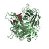
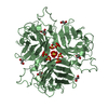
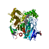
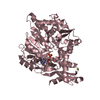
| Title | CRYSTAL STRUCTURE OF THE RECEPTOR-BINDING DOMAIN OF ADENOVIRUS TYPE 5 FIBER PROTEIN AT 1.7 ANGSTROMS RESOLUTION | ||||||
 Components Components | ADENOVIRUS TYPE 5 FIBER PROTEIN | ||||||
 Keywords Keywords | CELL RECEPTOR RECOGNITION | ||||||
| Function / homology |  Function and homology information Function and homology informationviral capsid, fiber / adhesion receptor-mediated virion attachment to host cell / cell adhesion / symbiont entry into host cell / host cell nucleus Similarity search - Function | ||||||
| Biological species |   Human adenovirus 5 Human adenovirus 5 | ||||||
| Method |  X-RAY DIFFRACTION / Resolution: 1.7 Å X-RAY DIFFRACTION / Resolution: 1.7 Å | ||||||
 Authors Authors | Xia, D. / Henry, L.J. / Gerard, R.D. / Deisenhofer, J. | ||||||
 Citation Citation | Journal: Structure / Year: 1994 Title: Crystal structure of the receptor-binding domain of adenovirus type 5 fiber protein at 1.7 A resolution. Authors: Xia, D. / Henry, L.J. / Gerard, R.D. / Deisenhofer, J. #1: Journal: J.Virol. / Year: 1994Title: Characterization of the Knob Domain of the Adenovirus Type 5 Fiber Protein Expressed in Escherichia Coli Authors: Henry, L.J. / Xia, D. / Wilke, M.E. / Deisenhofer, J. / Gerard, R.D. #2: Journal: J.Mol.Biol. / Year: 1994Title: Structure of Adenovirus Fiber: Morphology of Single Fibers Authors: Ruigrok, R.W.H. / Barge, A. / Albiges-Rizo, C. / Dayan, S. | ||||||
| History |
|
- Structure visualization
| Structure viewer | Molecule: MolmilJmol/JSmol |
|---|
- Downloads & links
-Download
| PDBx/mmCIF format | 1knb.cif.gz | 61.5 KB | Display | PDBx/mmCIF format |
|---|---|---|---|---|
| PDB format | pdb1knb.ent.gz | 46.1 KB | Display | PDB format |
| PDBx/mmJSON format | 1knb.json.gz | Tree view | PDBx/mmJSON format | |
| Others |  Other downloads Other downloads |
-Validation report
| Arichive directory | https://data.pdbj.org/pub/pdb/validation_reports/kn/1knbftp://data.pdbj.org/pub/pdb/validation_reports/kn/1knb | HTTPS FTP |
|---|
-Related structure data
| Similar structure data |
|---|


-Links
 PDBj
PDBj
- Assembly
| Deposited unit | 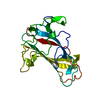
| ||||||||
|---|---|---|---|---|---|---|---|---|---|
| 1 | 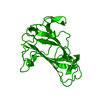
| ||||||||
| Unit cell |
| ||||||||
| Atom site foot note | 1: CIS PROLINE - PRO 447 |
-Components
| #1: Protein | Mass: 21256.695 Da / Num. of mol.: 1 Source method: isolated from a genetically manipulated source Source: (gene. exp.) Human adenovirus 5 / Genus: Mastadenovirus / Species: Human adenovirus C / Production host:  |
|---|---|
| #2: Water | ChemComp-HOH /  Mass: 18.015 Da / Num. of mol.: 145 / Source method: isolated from a natural source / Formula: H2O Mass: 18.015 Da / Num. of mol.: 145 / Source method: isolated from a natural source / Formula: H2O |
-Experimental details
-Experiment
| Experiment | Method: X-RAY DIFFRACTION |
|---|
- Sample preparation
| Crystal | Density Matthews: 2.53 Å3/Da / Density % sol: 51.33 % | ||||||||||||||||||||||||||||||||||||||||
|---|---|---|---|---|---|---|---|---|---|---|---|---|---|---|---|---|---|---|---|---|---|---|---|---|---|---|---|---|---|---|---|---|---|---|---|---|---|---|---|---|---|
| Crystal grow | *PLUS Method: vapor diffusion, hanging drop | ||||||||||||||||||||||||||||||||||||||||
| Components of the solutions | *PLUS
|
-Data collection
| Radiation | Scattering type: x-ray |
|---|---|
| Radiation wavelength | Relative weight: 1 |
| Reflection | Resolution: 1.7→12 Å / Num. obs: 17633 / % possible obs: 73.9 % / Observed criterion σ(I): 2 |
- Processing
| Software |
| ||||||||||||||||||||||||||||||||||||||||||||||||||||||||||||
|---|---|---|---|---|---|---|---|---|---|---|---|---|---|---|---|---|---|---|---|---|---|---|---|---|---|---|---|---|---|---|---|---|---|---|---|---|---|---|---|---|---|---|---|---|---|---|---|---|---|---|---|---|---|---|---|---|---|---|---|---|---|
| Refinement | Rfactor Rwork: 0.158 / Rfactor obs: 0.158 / Highest resolution: 1.7 Å Details: IN THE MONOMER STRUCTURE, THERE ARE TWO REGIONS WHERE B FACTORS ARE HIGH. ONE REGION IS FROM 396 - 397 WHICH IS LOCATED AT THE N-TERMINUS. THE OTHER REGION IS BETWEEN BETA STRANDS H AND I FOR RESIDUES 540 - 546. | ||||||||||||||||||||||||||||||||||||||||||||||||||||||||||||
| Refinement step | Cycle: LAST / Highest resolution: 1.7 Å
| ||||||||||||||||||||||||||||||||||||||||||||||||||||||||||||
| Refine LS restraints |
|
