 Movie
Movie Controller


+ Open data

- Basic information
| Entry | Database: PDB / ID: 1tme | ||||||
|---|---|---|---|---|---|---|---|
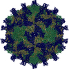
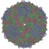
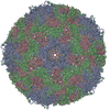
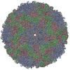
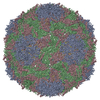
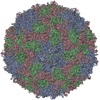
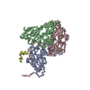
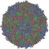
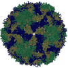
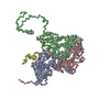
| Title | THREE-DIMENSIONAL STRUCTURE OF THEILER VIRUS | ||||||
 Components Components |
| ||||||
 Keywords Keywords | VIRUS / Icosahedral virus | ||||||
| Function / homology |  Function and homology information Function and homology informationhost cell nucleolus / picornain 3C / T=pseudo3 icosahedral viral capsid / host cell cytoplasmic vesicle membrane / channel activity / monoatomic ion transmembrane transport / symbiont-mediated suppression of host cytoplasmic pattern recognition receptor signaling pathway via inhibition of IRF3 activity / RNA helicase activity / RNA helicase / symbiont-mediated suppression of host gene expression ...host cell nucleolus / picornain 3C / T=pseudo3 icosahedral viral capsid / host cell cytoplasmic vesicle membrane / channel activity / monoatomic ion transmembrane transport / symbiont-mediated suppression of host cytoplasmic pattern recognition receptor signaling pathway via inhibition of IRF3 activity / RNA helicase activity / RNA helicase / symbiont-mediated suppression of host gene expression / symbiont-mediated activation of host autophagy / RNA-directed RNA polymerase / cysteine-type endopeptidase activity / viral RNA genome replication / RNA-directed RNA polymerase activity / symbiont entry into host cell / DNA-templated transcription / virion attachment to host cell / structural molecule activity / ATP hydrolysis activity / proteolysis / RNA binding / zinc ion binding / ATP binding Similarity search - Function | ||||||
| Biological species |  Theiler's encephalomyelitis virus Theiler's encephalomyelitis virus | ||||||
| Method |  X-RAY DIFFRACTION / Resolution: 2.8 Å X-RAY DIFFRACTION / Resolution: 2.8 Å | ||||||
 Authors Authors | Grant, R.A. / Filman, D.J. / Hogle, J.M. | ||||||
 Citation Citation | Journal: Proc.Natl.Acad.Sci.USA / Year: 1992 Title: Three-dimensional structure of Theiler virus. Authors: Grant, R.A. / Filman, D.J. / Fujinami, R.S. / Icenogle, J.P. / Hogle, J.M. #1: Journal: Embo J. / Year: 1989Title: Structural Factors that Control Conformational Transitions and Serotype Specificity in Type 3 Poliovirus Authors: Filman, D.J. / Syed, R. / Chow, M. / Macadam, A.J. / Minor, P.D. / Hogle, J.M. | ||||||
| History |
| ||||||
| Remark 285 | THE ENTRY PRESENTED HERE DOES NOT CONTAIN THE COMPLETE CRYSTAL ASYMMETRIC UNIT. IN ADDITION, THE ...THE ENTRY PRESENTED HERE DOES NOT CONTAIN THE COMPLETE CRYSTAL ASYMMETRIC UNIT. IN ADDITION, THE COORDINATES ARE NOT PRESENTED IN THE STANDARD CRYSTAL FRAME. IN ORDER TO GENERATE THE FULL CRYSTAL AU, APPLY THE FOLLOWING TRANSFORMATION MATRIX OR MATRICES AND SELECTED BIOMT RECORDS TO THE COORDINATES, AS SHOWN BELOW. X0 1 1.000000 0.000000 0.000000 0.00000 X0 2 0.000000 1.000000 0.000000 0.00000 X0 3 0.000000 0.000000 1.000000 0.00000 CRYSTAL AU = (X0) * (BIOMT 1-5,11-15,21-30,41-50) * CHAINS 1,2,3,4 |
- Structure visualization
| Structure viewer | Molecule: MolmilJmol/JSmol |
|---|
- Downloads & links
-Download
| PDBx/mmCIF format | 1tme.cif.gz | 152.5 KB | Display | PDBx/mmCIF format |
|---|---|---|---|---|
| PDB format | pdb1tme.ent.gz | 115.5 KB | Display | PDB format |
| PDBx/mmJSON format | 1tme.json.gz | Tree view | PDBx/mmJSON format | |
| Others |  Other downloads Other downloads |
-Validation report
| Arichive directory | https://data.pdbj.org/pub/pdb/validation_reports/tm/1tmeftp://data.pdbj.org/pub/pdb/validation_reports/tm/1tme | HTTPS FTP |
|---|
-Related structure data
| Similar structure data |
|---|
-Links
 PDBj
PDBj

- Assembly
| Deposited unit | 
| ||||||||
|---|---|---|---|---|---|---|---|---|---|
| 1 | x 60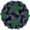
| ||||||||
| 2 |
| ||||||||
| 3 | x 5
| ||||||||
| 4 | x 6
| ||||||||
| 5 | 
| ||||||||
| 6 | x 30
| ||||||||
| Unit cell |
| ||||||||
| Atom site foot note | 1: CIS PROLINE - PRO 1 105 / 2: CIS PROLINE - PRO 2 85 3: SOLVENT MOLECULES WITH OCCUPANCIES SIGNIFICANTLY GREATER THAN 1.0 ARE SUSPECTED TO BE ANION OR CATION SITES. | ||||||||
| Symmetry | Point symmetry: (Hermann–Mauguin notation: 532 / Schoenflies symbol: I (icosahedral)) |
-Components
| #1: Protein | Mass: 30385.385 Da / Num. of mol.: 1 Source method: isolated from a genetically manipulated source Source: (gene. exp.) Theiler's encephalomyelitis virus (STRAIN DA)Genus: Cardiovirus / Species: Theilovirus / Strain: DA / Organ: BEAN / References: UniProt: P13899 |
|---|---|
| #2: Protein | Mass: 29544.609 Da / Num. of mol.: 1 Source method: isolated from a genetically manipulated source Source: (gene. exp.) Theiler's encephalomyelitis virus (STRAIN DA)Genus: Cardiovirus / Species: Theilovirus / Strain: DA / Organ: BEAN / References: UniProt: P13899 |
| #3: Protein | Mass: 25824.186 Da / Num. of mol.: 1 Source method: isolated from a genetically manipulated source Source: (gene. exp.) Theiler's encephalomyelitis virus (STRAIN DA)Genus: Cardiovirus / Species: Theilovirus / Strain: DA / Organ: BEAN / References: UniProt: P13899 |
| #4: Protein | Mass: 7107.480 Da / Num. of mol.: 1 Source method: isolated from a genetically manipulated source Source: (gene. exp.) Theiler's encephalomyelitis virus (STRAIN DA)Genus: Cardiovirus / Species: Theilovirus / Strain: DA / Organ: BEAN / References: UniProt: P13899 |
| #5: Water | ChemComp-HOH /  Mass: 18.015 Da / Num. of mol.: 327 / Source method: isolated from a natural source / Formula: H2O Mass: 18.015 Da / Num. of mol.: 327 / Source method: isolated from a natural source / Formula: H2O |
| Nonpolymer details | SOLVENT MOLECULES WITH OCCUPANCIE |
-Experimental details
-Experiment
| Experiment | Method: X-RAY DIFFRACTION |
|---|
- Sample preparation
| Crystal grow | *PLUS Temperature: 4 ℃ / Method: microdialysis / PH range low: 7.3 / PH range high: 7 | ||||||||||||||||||||||||||||
|---|---|---|---|---|---|---|---|---|---|---|---|---|---|---|---|---|---|---|---|---|---|---|---|---|---|---|---|---|---|
| Components of the solutions | *PLUS
|
-Data collection
| Reflection | *PLUS Highest resolution: 2.8 Å / Num. obs: 473861 / Num. measured all: 1154291 / Rmerge(I) obs: 0.2 |
|---|
- Processing
| Software | Name: REAL-SPACE / Version: REFINEMENT / Classification: refinement | ||||||||||||||||||||||||||||||||||||||||||||||||||||||||||||
|---|---|---|---|---|---|---|---|---|---|---|---|---|---|---|---|---|---|---|---|---|---|---|---|---|---|---|---|---|---|---|---|---|---|---|---|---|---|---|---|---|---|---|---|---|---|---|---|---|---|---|---|---|---|---|---|---|---|---|---|---|---|
| Refinement | Resolution: 2.8→30 Å Details: THE ELECTRON DENSITY WAS INCONSISTENT WITH THE PREDICTED AMINO ACID SEQUENCE DERIVED FROM THE GENE SEQUENCE OF THE DA STRAIN OF TMEV AT THREE SITES. THE STRUCTURE PRESENTED IN THIS ENTRY ...Details: THE ELECTRON DENSITY WAS INCONSISTENT WITH THE PREDICTED AMINO ACID SEQUENCE DERIVED FROM THE GENE SEQUENCE OF THE DA STRAIN OF TMEV AT THREE SITES. THE STRUCTURE PRESENTED IN THIS ENTRY REFLECTS THE ELECTRON DENSITY AT THESE SITES. RESIDUE 214 OF VP1 WAS BUILT AS GLY. THIS RESIDUE HAS BEEN REPORTED TO BE EITHER LEU (Y.OHARA,S.STEIN,J.FU,L.STILLMAN, L.KLAMAN,R.P.ROOS (1988) VIROLOGY 164, 244-255) OR TRP (A.ZURBRIGGEN,J.M.HOGLE,R.S.FUJINAMI (1989) J.EXP.MED. 170, 2037-2049). SURROUNDING PORTIONS OF THE STRUCTURE PACK TOO CLOSELY TO PERMIT THIS RESIDUE TO HAVE A SIDE CHAIN AND NO SIDE CHAIN DENSITY IS VISIBLE IN THE MAP. RESIDUES 202 AND 230 OF VP3 ARE REPORTED TO BE ALA IN THE DATABASE BUT IN BOTH CASES THE ELECTRON DENSITY SUGGEST A LARGER SIDE CHAIN, EITHER THR OR VAL. A THR SIDE CHAIN WAS BUILT INTO POSITION 202 AND A VAL INTO POSITION 230. IN BOTH CASES THE CHANGE IN ASSIGNMENT IS CONSISTENT WITH THE LOCAL ENVIRONMENT OF THE SIDE CHAIN AND WITH THE SEQUENCE OF THE BEAN AND GDVII STRAINS OF TMEV. THE PRESENCE OF THR AT POSITION 202 HAS BEEN CONFIRMED BY THE SEQUENCING OF THE DA SEED STOCK (UNPUBLISHED DATA). ANOTHER DIFFERENCE BETWEEN THE SEQUENCE DATA BASE AND THE STRUCTURE PRESENTED IN THIS ENTRY IS AT RESIDUE 266 OF VP2 WHERE THE RESIDUE IN THE ENTRY WAS REFINED AS GLY INSTEAD OF ALA. THIS RESIDUE, THE CARBOXYL TERMINUS OF VP2, IS PARTLY DISORDERED AND NO SIDE CHAIN DENSITY WAS VISIBLE IN THE MAP.
| ||||||||||||||||||||||||||||||||||||||||||||||||||||||||||||
| Refinement step | Cycle: LAST / Resolution: 2.8→30 Å
| ||||||||||||||||||||||||||||||||||||||||||||||||||||||||||||
| Refine LS restraints |
| ||||||||||||||||||||||||||||||||||||||||||||||||||||||||||||
| Software | *PLUS Name: 'PSEUDO-REAL SPACE PROCEDURE' / Classification: refinement | ||||||||||||||||||||||||||||||||||||||||||||||||||||||||||||
| Refinement | *PLUS Highest resolution: 2.8 Å / Lowest resolution: 30 Å / Num. reflection obs: 473961 / Rfactor obs: 0.3 | ||||||||||||||||||||||||||||||||||||||||||||||||||||||||||||
| Solvent computation | *PLUS | ||||||||||||||||||||||||||||||||||||||||||||||||||||||||||||
| Displacement parameters | *PLUS |
