 Movie
Movie Controller


+ Open data

- Basic information
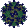
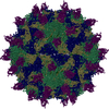
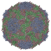
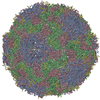
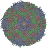
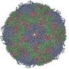
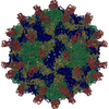
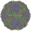
| Entry | Database: PDB / ID: 6gv4 | |||||||||||||||
|---|---|---|---|---|---|---|---|---|---|---|---|---|---|---|---|---|
| Title | High-resolution Cryo-EM of Fab-labeled human parechovirus 3 | |||||||||||||||
 Components Components |
| |||||||||||||||
 Keywords Keywords | VIRUS / human parechovirus / antibody / RNA | |||||||||||||||
| Function / homology |  Function and homology information Function and homology informationhost cell nucleolus / T=pseudo3 icosahedral viral capsid / host cell cytoplasmic vesicle membrane / ribonucleoside triphosphate phosphatase activity / channel activity / monoatomic ion transmembrane transport / RNA helicase activity / cysteine-type endopeptidase activity / viral RNA genome replication / RNA-directed RNA polymerase activity ...host cell nucleolus / T=pseudo3 icosahedral viral capsid / host cell cytoplasmic vesicle membrane / ribonucleoside triphosphate phosphatase activity / channel activity / monoatomic ion transmembrane transport / RNA helicase activity / cysteine-type endopeptidase activity / viral RNA genome replication / RNA-directed RNA polymerase activity / symbiont entry into host cell / DNA-templated transcription / virion attachment to host cell / structural molecule activity / proteolysis / RNA binding / ATP binding / metal ion binding Similarity search - Function | |||||||||||||||
| Biological species |  Homo sapiens (human) Homo sapiens (human) Human parechovirus 3 Human parechovirus 3 | |||||||||||||||
| Method | ELECTRON MICROSCOPY / single particle reconstruction / cryo EM / Resolution: 2.8 Å | |||||||||||||||
 Authors Authors | Domanska, A. / Flatt, J.W. / Jukonen, J.J.J. / Geraets, J.A. / Butcher, S.J. | |||||||||||||||
| Funding support |  Finland, 4items Finland, 4items
| |||||||||||||||
 Citation Citation | Journal: J Virol / Year: 2019 Title: A 2.8-Angstrom-Resolution Cryo-Electron Microscopy Structure of Human Parechovirus 3 in Complex with Fab from a Neutralizing Antibody. Authors: Aušra Domanska / Justin W Flatt / Joonas J J Jukonen / James A Geraets / Sarah J Butcher / Abstract: Human parechovirus 3 (HPeV3) infection is associated with sepsis characterized by significant immune activation and subsequent tissue damage in neonates. Strategies to limit infection have been ...Human parechovirus 3 (HPeV3) infection is associated with sepsis characterized by significant immune activation and subsequent tissue damage in neonates. Strategies to limit infection have been unsuccessful due to inadequate molecular diagnostic tools for early detection and the lack of a vaccine or specific antiviral therapy. Toward the latter, we present a 2.8-Å-resolution structure of HPeV3 in complex with fragments from a neutralizing human monoclonal antibody, AT12-015, using cryo-electron microscopy (cryo-EM) and image reconstruction. Modeling revealed that the epitope extends across neighboring asymmetric units with contributions from capsid proteins VP0, VP1, and VP3. Antibody decoration was found to block binding of HPeV3 to cultured cells. Additionally, at high resolution, it was possible to model a stretch of RNA inside the virion and, from this, identify the key features that drive and stabilize protein-RNA association during assembly. Human parechovirus 3 (HPeV3) is receiving increasing attention as a prevalent cause of sepsis-like symptoms in neonates, for which, despite the severity of disease, there are no effective treatments available. Structural and molecular insights into virus neutralization are urgently needed, especially as clinical cases are on the rise. Toward this goal, we present the first structure of HPeV3 in complex with fragments from a neutralizing monoclonal antibody. At high resolution, it was possible to precisely define the epitope that, when targeted, prevents virions from binding to cells. Such an atomic-level description is useful for understanding host-pathogen interactions and viral pathogenesis mechanisms and for finding potential cures for infection and disease. | |||||||||||||||
| History |
|
- Structure visualization
| Movie |
Movie viewer |
|---|---|
| Structure viewer | Molecule: MolmilJmol/JSmol |
- Downloads & links
-Download
| PDBx/mmCIF format | 6gv4.cif.gz | 306.9 KB | Display | PDBx/mmCIF format |
|---|---|---|---|---|
| PDB format | pdb6gv4.ent.gz | 242.7 KB | Display | PDB format |
| PDBx/mmJSON format | 6gv4.json.gz | Tree view | PDBx/mmJSON format | |
| Others |  Other downloads Other downloads |
-Validation report
| Arichive directory | https://data.pdbj.org/pub/pdb/validation_reports/gv/6gv4ftp://data.pdbj.org/pub/pdb/validation_reports/gv/6gv4 | HTTPS FTP |
|---|
-Related structure data
| Related structure data |  0069MC M: map data used to model this data C: citing same article ( |
|---|---|
| Similar structure data | |
| EM raw data | EMPIAR-10983 (Title: High-resolution Cryo-EM of Fab-labeled human parechovirus 3 Data size: 3.3 TB Data #1: Unaligned multi-frame micrographs of HPeV3-fab complex [micrographs - multiframe]) |


-Links
 PDBj
PDBj


- Assembly
| Deposited unit | 
|
|---|---|
| 1 | x 60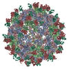
|
-Components
-RNA chain , 1 types, 1 molecules D
| #1: RNA chain | Mass: 2505.489 Da / Num. of mol.: 1 / Source method: isolated from a natural source / Details: RNA / Source: (natural) Human parechovirus 3 / Cell line: HT29 / Organ: colon adenocarcinoma |
|---|
-Protein , 3 types, 3 molecules ABC
| #2: Protein | Mass: 31770.135 Da / Num. of mol.: 1 / Source method: isolated from a natural source / Source: (natural) Human parechovirus 3 / Cell line: HT29 / Organ: colon adenocarcinoma / References: UniProt: Q8BES5 |
|---|---|
| #3: Protein | Mass: 25913.127 Da / Num. of mol.: 1 / Source method: isolated from a natural source / Source: (natural) Human parechovirus 3 / Cell line: HT29 / Organ: colon adenocarcinoma / References: UniProt: Q8BES5 |
| #4: Protein | Mass: 28757.551 Da / Num. of mol.: 1 / Source method: isolated from a natural source / Details: polypeptide chain / Source: (natural) Human parechovirus 3 / Cell line: HT29 / Organ: colon adenocarcinoma / References: UniProt: Q8BES5, UniProt: A0A291FGQ4*PLUS |
-Antibody , 2 types, 2 molecules HL
| #5: Antibody | Mass: 13128.613 Da / Num. of mol.: 1 Source method: isolated from a genetically manipulated source Details: polypeptide chain / Source: (gene. exp.) Homo sapiens (human) / Cell line (production host): 293T / Production host: Homo sapiens (human) |
|---|---|
| #6: Antibody | Mass: 12339.752 Da / Num. of mol.: 1 Source method: isolated from a genetically manipulated source Details: polypeptide chain / Source: (gene. exp.) Homo sapiens (human) / Cell line (production host): 293T / Production host: Homo sapiens (human) |
-Details
| Has protein modification | Y |
|---|
-Experimental details
-Experiment
| Experiment | Method: ELECTRON MICROSCOPY |
|---|---|
| EM experiment | Aggregation state: PARTICLE / 3D reconstruction method: single particle reconstruction |
- Sample preparation
| Component |
| ||||||||||||||||||||||||
|---|---|---|---|---|---|---|---|---|---|---|---|---|---|---|---|---|---|---|---|---|---|---|---|---|---|
| Molecular weight | Value: 7.7 MDa / Experimental value: NO | ||||||||||||||||||||||||
| Source (natural) |
| ||||||||||||||||||||||||
| Source (recombinant) | Organism: Homo sapiens (human) | ||||||||||||||||||||||||
| Details of virus | Empty: NO / Enveloped: NO / Isolate: STRAIN / Type: VIRION | ||||||||||||||||||||||||
| Buffer solution | pH: 7.5 | ||||||||||||||||||||||||
| Buffer component |
| ||||||||||||||||||||||||
| Specimen | Conc.: 0.1 mg/ml / Embedding applied: NO / Shadowing applied: NO / Staining applied: NO / Vitrification applied: YES | ||||||||||||||||||||||||
| Specimen support | Details: ultrathin carbon-coated lacey 400-mesh copper grids (Ted Pella product #01824) Grid material: COPPER / Grid mesh size: 400 divisions/in. | ||||||||||||||||||||||||
| Vitrification | Instrument: HOMEMADE PLUNGER / Cryogen name: ETHANE / Chamber temperature: 295 K Details: We could not control humidity during plunging. It was ambient humidity. Blot for 1 s before plunging. |
- Electron microscopy imaging
| Experimental equipment |  Model: Titan Krios / Image courtesy: FEI Company |
|---|---|
| Microscopy | Model: FEI TITAN KRIOS |
| Electron gun | Electron source:  FIELD EMISSION GUN / Accelerating voltage: 300 kV / Illumination mode: FLOOD BEAM FIELD EMISSION GUN / Accelerating voltage: 300 kV / Illumination mode: FLOOD BEAM |
| Electron lens | Mode: BRIGHT FIELD / Nominal magnification: 75000 X / Nominal defocus max: 2500 nm / Nominal defocus min: 500 nm / Cs: 2.7 mm |
| Specimen holder | Cryogen: NITROGEN / Specimen holder model: FEI TITAN KRIOS AUTOGRID HOLDER |
| Image recording | Average exposure time: 1 sec. / Electron dose: 48 e/Å2 / Detector mode: INTEGRATING / Film or detector model: FEI FALCON II (4k x 4k) / Num. of grids imaged: 2 / Num. of real images: 6541 |
| Image scans | Movie frames/image: 18 / Used frames/image: 2-17 |
- Processing
| EM software |
| ||||||||||||||||||||||||||||||||||||||||||||||||||
|---|---|---|---|---|---|---|---|---|---|---|---|---|---|---|---|---|---|---|---|---|---|---|---|---|---|---|---|---|---|---|---|---|---|---|---|---|---|---|---|---|---|---|---|---|---|---|---|---|---|---|---|
| CTF correction | Details: GCTF was used to estimate ctf / Type: PHASE FLIPPING AND AMPLITUDE CORRECTION | ||||||||||||||||||||||||||||||||||||||||||||||||||
| Particle selection | Num. of particles selected: 217212 Details: automatic particle selection in RELION using template generated from manually selected particles | ||||||||||||||||||||||||||||||||||||||||||||||||||
| Symmetry | Point symmetry: I (icosahedral) | ||||||||||||||||||||||||||||||||||||||||||||||||||
| 3D reconstruction | Resolution: 2.8 Å / Resolution method: FSC 0.143 CUT-OFF / Num. of particles: 74927 / Num. of class averages: 3 / Symmetry type: POINT | ||||||||||||||||||||||||||||||||||||||||||||||||||
| Atomic model building | Protocol: OTHER / Space: REAL Details: Initial model was generated in I-TASSER and SWISSMODEL using 4z92 and 4udf as reference. Initial rigid fit of the model to the map was done in UCSF Chimera. Model refinement was done in Coot and MDFF. |
