 Movie
Movie Controller


+ Open data

- Basic information
| Entry | Database: PDB / ID: 6egx | |||||||||
|---|---|---|---|---|---|---|---|---|---|---|
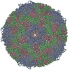
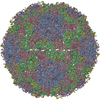
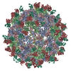
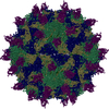
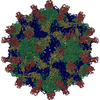
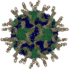
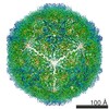
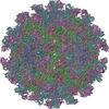
| Title | Sacbrood virus of honeybee - expansion state I | |||||||||
 Components Components |
| |||||||||
 Keywords Keywords | VIRUS / empty particle / icosahedral virus particle / iflavirus / expanded particle | |||||||||
| Function / homology |  Function and homology information Function and homology informationhost cell membrane / viral capsid / RNA helicase activity / cysteine-type endopeptidase activity / viral RNA genome replication / RNA-directed RNA polymerase activity / DNA-templated transcription / structural molecule activity / proteolysis / RNA binding / ATP binding Similarity search - Function | |||||||||
| Biological species |  Sacbrood virus Sacbrood virus | |||||||||
| Method | ELECTRON MICROSCOPY / single particle reconstruction / cryo EM / Resolution: 4.06 Å | |||||||||
 Authors Authors | Plevka, P. / Prochazkova, M. | |||||||||
| Funding support |  Czech Republic, 2items Czech Republic, 2items
| |||||||||
 Citation Citation | Journal: Proc Natl Acad Sci U S A / Year: 2018 Title: Virion structure and genome delivery mechanism of sacbrood honeybee virus. Authors: Michaela Procházková / Tibor Füzik / Karel Škubník / Jana Moravcová / Zorica Ubiparip / Antonín Přidal / Pavel Plevka / Abstract: Infection by sacbrood virus (SBV) from the family Iflaviridae is lethal to honey bee larvae but only rarely causes the collapse of honey bee colonies. Despite the negative effect of SBV on honey ...Infection by sacbrood virus (SBV) from the family Iflaviridae is lethal to honey bee larvae but only rarely causes the collapse of honey bee colonies. Despite the negative effect of SBV on honey bees, the structure of its particles and mechanism of its genome delivery are unknown. Here we present the crystal structure of SBV virion and show that it contains 60 copies of a minor capsid protein (MiCP) attached to the virion surface. No similar MiCPs have been previously reported in any of the related viruses from the order Picornavirales. The location of the MiCP coding sequence within the SBV genome indicates that the MiCP evolved from a C-terminal extension of a major capsid protein by the introduction of a cleavage site for a virus protease. The exposure of SBV to acidic pH, which the virus likely encounters during cell entry, induces the formation of pores at threefold and fivefold axes of the capsid that are 7 Å and 12 Å in diameter, respectively. This is in contrast to vertebrate picornaviruses, in which the pores along twofold icosahedral symmetry axes are currently considered the most likely sites for genome release. SBV virions lack VP4 subunits that facilitate the genome delivery of many related dicistroviruses and picornaviruses. MiCP subunits induce liposome disruption in vitro, indicating that they are functional analogs of VP4 subunits and enable the virus genome to escape across the endosome membrane into the cell cytoplasm. | |||||||||
| History |
|
- Structure visualization
| Movie |
Movie viewer |
|---|---|
| Structure viewer | Molecule: MolmilJmol/JSmol |
- Downloads & links
-Download
| PDBx/mmCIF format | 6egx.cif.gz | 159.3 KB | Display | PDBx/mmCIF format |
|---|---|---|---|---|
| PDB format | pdb6egx.ent.gz | 121.5 KB | Display | PDB format |
| PDBx/mmJSON format | 6egx.json.gz | Tree view | PDBx/mmJSON format | |
| Others |  Other downloads Other downloads |
-Validation report
| Arichive directory | https://data.pdbj.org/pub/pdb/validation_reports/eg/6egxftp://data.pdbj.org/pub/pdb/validation_reports/eg/6egx | HTTPS FTP |
|---|
-Related structure data
| Related structure data |  3866MC 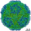 3863C 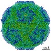 3865C  3867C  3881C 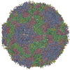 5lsfC  5oypC 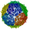 6egvC  6eh1C  6eiwC M: map data used to model this data C: citing same article ( |
|---|---|
| Similar structure data |


-Links
 PDBj
PDBj
- Assembly
| Deposited unit | 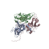
|
|---|---|
| 1 | x 60
|
| 2 |
|
| 3 | x 5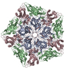
|
| 4 | x 6
|
| 5 | 
|
| Symmetry | Point symmetry: (Schoenflies symbol: I (icosahedral)) |
-Components
| #1: Protein | Mass: 26151.500 Da / Num. of mol.: 1 / Source method: isolated from a natural source / Source: (natural) Sacbrood virus / Plasmid details: Petrusov, CZ / References: UniProt: A0A223DN59, UniProt: Q9WCE9*PLUS |
|---|---|
| #2: Protein | Mass: 22563.734 Da / Num. of mol.: 1 / Source method: isolated from a natural source / Source: (natural) Sacbrood virus / Plasmid details: Petrusov, CZ / References: UniProt: A0A223DN66 |
| #3: Protein | Mass: 30701.373 Da / Num. of mol.: 1 / Source method: isolated from a natural source / Source: (natural) Sacbrood virus / Plasmid details: Petrusov, CZ / References: UniProt: A0A2I6HDZ6 |
| #4: Protein/peptide | Mass: 3107.398 Da / Num. of mol.: 1 / Source method: isolated from a natural source / Source: (natural) Sacbrood virus / Plasmid details: Petrusov, CZ / References: UniProt: Q9IGK7 |
-Experimental details
-Experiment
| Experiment | Method: ELECTRON MICROSCOPY |
|---|---|
| EM experiment | Aggregation state: PARTICLE / 3D reconstruction method: single particle reconstruction |
- Sample preparation
| Component | Name: Sacbrood virus / Type: VIRUS / Details: Isolated from honeybee pupae / Entity ID: all / Source: NATURAL | |||||||||||||||||||||||||
|---|---|---|---|---|---|---|---|---|---|---|---|---|---|---|---|---|---|---|---|---|---|---|---|---|---|---|
| Molecular weight | Experimental value: NO | |||||||||||||||||||||||||
| Source (natural) | Organism: Sacbrood virus | |||||||||||||||||||||||||
| Details of virus | Empty: YES / Enveloped: NO / Isolate: STRAIN / Type: VIRION | |||||||||||||||||||||||||
| Natural host | Organism: Apis mellifera | |||||||||||||||||||||||||
| Virus shell | Name: capsid / Diameter: 274.96 nm / Triangulation number (T number): 3 | |||||||||||||||||||||||||
| Buffer solution | pH: 5.8 | |||||||||||||||||||||||||
| Buffer component |
| |||||||||||||||||||||||||
| Specimen | Conc.: 15 mg/ml / Embedding applied: NO / Shadowing applied: NO / Staining applied: NO / Vitrification applied: YES | |||||||||||||||||||||||||
| Specimen support | Grid material: COPPER / Grid mesh size: 300 divisions/in. / Grid type: Quantifoil R2/1 | |||||||||||||||||||||||||
| Vitrification | Instrument: FEI VITROBOT MARK II / Cryogen name: ETHANE / Humidity: 70 % / Chamber temperature: 298 K |
- Electron microscopy imaging
| Experimental equipment |  Model: Titan Krios / Image courtesy: FEI Company |
|---|---|
| Microscopy | Model: FEI TITAN KRIOS Details: Preliminar grid screening was performed manually on FEI Tecnai F20 (cryo stage). |
| Electron gun | Electron source:  FIELD EMISSION GUN / Accelerating voltage: 300 kV / Illumination mode: FLOOD BEAM FIELD EMISSION GUN / Accelerating voltage: 300 kV / Illumination mode: FLOOD BEAM |
| Electron lens | Mode: BRIGHT FIELD / Nominal magnification: 75000 X / Calibrated magnification: 74325 X / Nominal defocus max: 4000 nm / Nominal defocus min: 1000 nm / Cs: 2.7 mm / C2 aperture diameter: 100 µm / Alignment procedure: COMA FREE |
| Specimen holder | Cryogen: NITROGEN / Specimen holder model: FEI TITAN KRIOS AUTOGRID HOLDER |
| Image recording | Average exposure time: 1 sec. / Electron dose: 21 e/Å2 / Detector mode: COUNTING / Film or detector model: FEI FALCON II (4k x 4k) / Num. of grids imaged: 1 |
| Image scans | Width: 4096 / Height: 4096 / Movie frames/image: 16 / Used frames/image: 2-16 |
- Processing
| Software | Name: REFMAC / Version: 5.8.0222 / Classification: refinement | ||||||||||||||||||||||||||||||||||||||||||||||||||||||||||||||||||||||||||||||||||||||||||||||||||||||||||
|---|---|---|---|---|---|---|---|---|---|---|---|---|---|---|---|---|---|---|---|---|---|---|---|---|---|---|---|---|---|---|---|---|---|---|---|---|---|---|---|---|---|---|---|---|---|---|---|---|---|---|---|---|---|---|---|---|---|---|---|---|---|---|---|---|---|---|---|---|---|---|---|---|---|---|---|---|---|---|---|---|---|---|---|---|---|---|---|---|---|---|---|---|---|---|---|---|---|---|---|---|---|---|---|---|---|---|---|
| EM software |
| ||||||||||||||||||||||||||||||||||||||||||||||||||||||||||||||||||||||||||||||||||||||||||||||||||||||||||
| CTF correction | Type: PHASE FLIPPING AND AMPLITUDE CORRECTION | ||||||||||||||||||||||||||||||||||||||||||||||||||||||||||||||||||||||||||||||||||||||||||||||||||||||||||
| Particle selection | Num. of particles selected: 51000 Details: Full virions were selected together with empty particles from the same set of micrographs. Subsequent 2D classification sorted the particles of two types. Full and empty particles were ...Details: Full virions were selected together with empty particles from the same set of micrographs. Subsequent 2D classification sorted the particles of two types. Full and empty particles were further processed separately. Empty particles classified in two types - expansion state I and II. | ||||||||||||||||||||||||||||||||||||||||||||||||||||||||||||||||||||||||||||||||||||||||||||||||||||||||||
| 3D reconstruction | Resolution: 4.06 Å / Resolution method: FSC 0.143 CUT-OFF / Num. of particles: 2341 / Algorithm: FOURIER SPACE / Num. of class averages: 1 / Symmetry type: POINT | ||||||||||||||||||||||||||||||||||||||||||||||||||||||||||||||||||||||||||||||||||||||||||||||||||||||||||
| Atomic model building | B value: 114.26 / Protocol: RIGID BODY FIT / Space: REAL / Target criteria: R-factor | ||||||||||||||||||||||||||||||||||||||||||||||||||||||||||||||||||||||||||||||||||||||||||||||||||||||||||
| Refinement | Resolution: 4.06→4.06 Å / Cor.coef. Fo:Fc: 0.9 / SU B: 29.687 / SU ML: 0.365 / ESU R: 0.178 Stereochemistry target values: MAXIMUM LIKELIHOOD WITH PHASES Details: HYDROGENS HAVE BEEN ADDED IN THE RIDING POSITIONS
| ||||||||||||||||||||||||||||||||||||||||||||||||||||||||||||||||||||||||||||||||||||||||||||||||||||||||||
| Solvent computation | Ion probe radii: 0.8 Å / Shrinkage radii: 0.8 Å / VDW probe radii: 1.2 Å / Solvent model: MASK | ||||||||||||||||||||||||||||||||||||||||||||||||||||||||||||||||||||||||||||||||||||||||||||||||||||||||||
| Displacement parameters | Biso mean: 117.829 Å2
| ||||||||||||||||||||||||||||||||||||||||||||||||||||||||||||||||||||||||||||||||||||||||||||||||||||||||||
| Refinement step | Cycle: 1 / Total: 5819 | ||||||||||||||||||||||||||||||||||||||||||||||||||||||||||||||||||||||||||||||||||||||||||||||||||||||||||
| Refine LS restraints |
|