 Movie
Movie Controller


+ Open data

- Basic information
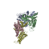
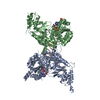
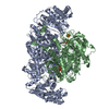
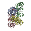
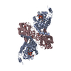
| Entry | Database: PDB / ID: 1jey | ||||||
|---|---|---|---|---|---|---|---|
| Title | Crystal Structure of the Ku heterodimer bound to DNA | ||||||
 Components Components |
| ||||||
 Keywords Keywords | DNA BINDING PROTEIN/DNA / double-strand DNA break repair / non-homologous end-joining / protein-nucleic acid complex / alpha/beta domain / beta barrel / helical C-terminal arm / SAP domain / DNA BINDING PROTEIN-DNA COMPLEX | ||||||
| Function / homology |  Function and homology information Function and homology informationKu70:Ku80 complex / negative regulation of t-circle formation / DNA end binding / small-subunit processome assembly / positive regulation of lymphocyte differentiation / DNA-dependent protein kinase complex / DNA-dependent protein kinase-DNA ligase 4 complex / nonhomologous end joining complex / regulation of smooth muscle cell proliferation / cellular response to X-ray ...Ku70:Ku80 complex / negative regulation of t-circle formation / DNA end binding / small-subunit processome assembly / positive regulation of lymphocyte differentiation / DNA-dependent protein kinase complex / DNA-dependent protein kinase-DNA ligase 4 complex / nonhomologous end joining complex / regulation of smooth muscle cell proliferation / cellular response to X-ray / double-strand break repair via classical nonhomologous end joining / Cytosolic sensors of pathogen-associated DNA / nuclear telomere cap complex / IRF3-mediated induction of type I IFN / recombinational repair / regulation of telomere maintenance / protein localization to chromosome, telomeric region / U3 snoRNA binding / positive regulation of neurogenesis / cellular hyperosmotic salinity response / 2-LTR circle formation / hematopoietic stem cell proliferation / telomeric repeat DNA binding / DNA 3'-5' helicase / hematopoietic stem cell differentiation / 5'-deoxyribose-5-phosphate lyase activity / ATP-dependent activity, acting on DNA / telomere maintenance via telomerase / site of DNA damage / cellular response to leukemia inhibitory factor / neurogenesis / activation of innate immune response / telomere maintenance / cyclin binding / DNA-(apurinic or apyrimidinic site) lyase / DNA helicase activity / Nonhomologous End-Joining (NHEJ) / small-subunit processome / cellular response to gamma radiation / protein-DNA complex / double-strand break repair via nonhomologous end joining / enzyme activator activity / double-strand break repair / double-stranded DNA binding / scaffold protein binding / DNA recombination / secretory granule lumen / transcription regulator complex / ficolin-1-rich granule lumen / damaged DNA binding / chromosome, telomeric region / transcription cis-regulatory region binding / ribonucleoprotein complex / innate immune response / negative regulation of DNA-templated transcription / ubiquitin protein ligase binding / Neutrophil degranulation / DNA damage response / nucleolus / positive regulation of DNA-templated transcription / protein-containing complex binding / positive regulation of transcription by RNA polymerase II / ATP hydrolysis activity / protein-containing complex / DNA binding / RNA binding / extracellular region / nucleoplasm / ATP binding / membrane / nucleus / plasma membrane / cytosol Similarity search - Function | ||||||
| Biological species |  Homo sapiens (human) Homo sapiens (human) | ||||||
| Method |  X-RAY DIFFRACTION / SYNCHROTRON / MOLECULAR REPLACEMENT / Resolution: 2.5 Å X-RAY DIFFRACTION / SYNCHROTRON / MOLECULAR REPLACEMENT / Resolution: 2.5 Å | ||||||
 Authors Authors | Walker, J.R. / Corpina, R.A. / Goldberg, J. | ||||||
 Citation Citation | Journal: Nature / Year: 2001 Title: Structure of the Ku heterodimer bound to DNA and its implications for double-strand break repair. Authors: Walker, J.R. / Corpina, R.A. / Goldberg, J. | ||||||
| History |
|
- Structure visualization
| Structure viewer | Molecule: MolmilJmol/JSmol |
|---|
- Downloads & links
-Download
| PDBx/mmCIF format | 1jey.cif.gz | 250.7 KB | Display | PDBx/mmCIF format |
|---|---|---|---|---|
| PDB format | pdb1jey.ent.gz | 193.1 KB | Display | PDB format |
| PDBx/mmJSON format | 1jey.json.gz | Tree view | PDBx/mmJSON format | |
| Others |  Other downloads Other downloads |
-Validation report
| Arichive directory | https://data.pdbj.org/pub/pdb/validation_reports/je/1jeyftp://data.pdbj.org/pub/pdb/validation_reports/je/1jey | HTTPS FTP |
|---|
-Related structure data
| Related structure data |  1jeqSC S: Starting model for refinement C: citing same article ( |
|---|---|
| Similar structure data |


-Links
 PDBj
PDBj


- Assembly
| Deposited unit | 
| ||||||||
|---|---|---|---|---|---|---|---|---|---|
| 1 |
| ||||||||
| Unit cell |
|
-Components
| #1: DNA chain | Mass: 6506.188 Da / Num. of mol.: 1 / Source method: obtained synthetically |
|---|---|
| #2: DNA chain | Mass: 10325.685 Da / Num. of mol.: 1 / Source method: obtained synthetically |
| #3: Protein | Mass: 69945.039 Da / Num. of mol.: 1 Source method: isolated from a genetically manipulated source Source: (gene. exp.) Homo sapiens (human) / Gene: G22P1 / Plasmid: pFASTBAC HT / Cell line (production host): Sf9 / Production host:   Spodoptera frugiperda (fall armyworm) / References: UniProt: P12956 Spodoptera frugiperda (fall armyworm) / References: UniProt: P12956 |
| #4: Protein | Mass: 64190.543 Da / Num. of mol.: 1 / Fragment: residues 1-565 Source method: isolated from a genetically manipulated source Source: (gene. exp.) Homo sapiens (human) / Gene: XRCC5 / Plasmid: pFASTBAC HT / Cell line (production host): Sf9 / Production host: Spodoptera frugiperda (fall armyworm) / References: UniProt: P13010 |
| #5: Water | ChemComp-HOH /  Mass: 18.015 Da / Num. of mol.: 255 / Source method: isolated from a natural source / Formula: H2O Mass: 18.015 Da / Num. of mol.: 255 / Source method: isolated from a natural source / Formula: H2O |
-Experimental details
-Experiment
| Experiment | Method: X-RAY DIFFRACTION / Number of used crystals: 1 |
|---|
- Sample preparation
| Crystal | Density Matthews: 2.23 Å3/Da / Density % sol: 45 % | |||||||||||||||||||||||||||||||||||
|---|---|---|---|---|---|---|---|---|---|---|---|---|---|---|---|---|---|---|---|---|---|---|---|---|---|---|---|---|---|---|---|---|---|---|---|---|
| Crystal grow | Temperature: 295 K / Method: vapor diffusion, hanging drop / pH: 8 Details: 26% polyethylene glycol 350 monomethyl ether, 50 mM Tris-HCL pH 8.0, 5 mM magnesium chloride, VAPOR DIFFUSION, HANGING DROP, temperature 295K | |||||||||||||||||||||||||||||||||||
| Components of the solutions |
| |||||||||||||||||||||||||||||||||||
| Crystal grow | *PLUS Temperature: 22 ℃ | |||||||||||||||||||||||||||||||||||
| Components of the solutions | *PLUS
|
-Data collection
| Diffraction | Mean temperature: 100 K |
|---|---|
| Diffraction source | Source: SYNCHROTRON / Site: NSLS  / Beamline: X9A / Wavelength: 0.97921 Å / Beamline: X9A / Wavelength: 0.97921 Å |
| Detector | Type: MARRESEARCH / Detector: CCD / Date: Mar 7, 2001 |
| Radiation | Protocol: SINGLE WAVELENGTH / Monochromatic (M) / Laue (L): M / Scattering type: x-ray |
| Radiation wavelength | Wavelength: 0.97921 Å / Relative weight: 1 |
| Reflection | Resolution: 2.5→25 Å / Num. all: 46460 / Num. obs: 46176 / % possible obs: 96.4 % / Observed criterion σ(F): -3 / Observed criterion σ(I): -3 / Redundancy: 3.1 % / Biso Wilson estimate: 28.2 Å2 / Rmerge(I) obs: 0.065 / Net I/σ(I): 17.5 |
| Reflection shell | Resolution: 2.5→2.54 Å / Redundancy: 2.7 % / Rmerge(I) obs: 0.306 / Mean I/σ(I) obs: 3.6 / Num. unique all: 46385 / % possible all: 67.5 |
| Reflection | *PLUS Highest resolution: 2.5 Å / Num. obs: 46385 / Num. measured all: 141891 |
| Reflection shell | *PLUS % possible obs: 82.3 % / Rmerge(I) obs: 0.311 |
- Processing
| Software |
| ||||||||||||||||||||||||||||||||||||||||||||
|---|---|---|---|---|---|---|---|---|---|---|---|---|---|---|---|---|---|---|---|---|---|---|---|---|---|---|---|---|---|---|---|---|---|---|---|---|---|---|---|---|---|---|---|---|---|
| Refinement | Method to determine structure: MOLECULAR REPLACEMENT Starting model: PDB ENTRY 1JEQ Resolution: 2.5→24.74 Å / Rfactor Rfree error: 0.006 / Data cutoff high absF: 2533650.68 / Data cutoff low absF: 0 / Isotropic thermal model: RESTRAINED / Cross valid method: THROUGHOUT / σ(F): 0 / σ(I): 0 / Stereochemistry target values: Engh & Huber
| ||||||||||||||||||||||||||||||||||||||||||||
| Solvent computation | Solvent model: FLAT MODEL / Bsol: 34.46 Å2 / ksol: 0.36 e/Å3 | ||||||||||||||||||||||||||||||||||||||||||||
| Displacement parameters | Biso mean: 40.1 Å2
| ||||||||||||||||||||||||||||||||||||||||||||
| Refine analyze |
| ||||||||||||||||||||||||||||||||||||||||||||
| Refinement step | Cycle: LAST / Resolution: 2.5→24.74 Å
| ||||||||||||||||||||||||||||||||||||||||||||
| Refine LS restraints |
| ||||||||||||||||||||||||||||||||||||||||||||
| LS refinement shell | Resolution: 2.5→2.66 Å / Rfactor Rfree error: 0.019 / Total num. of bins used: 6
| ||||||||||||||||||||||||||||||||||||||||||||
| Xplor file |
| ||||||||||||||||||||||||||||||||||||||||||||
| Software | *PLUS Name: CNS / Version: 1 / Classification: refinement | ||||||||||||||||||||||||||||||||||||||||||||
| Refinement | *PLUS Lowest resolution: 25 Å / σ(F): 0 / % reflection Rfree: 5 % / Rfactor obs: 0.218 / Rfactor Rfree: 0.276 | ||||||||||||||||||||||||||||||||||||||||||||
| Solvent computation | *PLUS | ||||||||||||||||||||||||||||||||||||||||||||
| Displacement parameters | *PLUS Biso mean: 40.1 Å2 | ||||||||||||||||||||||||||||||||||||||||||||
| Refine LS restraints | *PLUS
| ||||||||||||||||||||||||||||||||||||||||||||
| LS refinement shell | *PLUS Rfactor Rfree: 0.345 / % reflection Rfree: 4.6 % / Rfactor Rwork: 0.261 |
