 Movie
Movie Controller


+ Open data

- Basic information
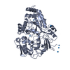
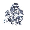
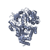
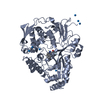
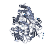
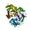
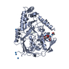
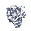
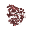
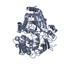
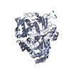
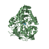
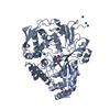
| Entry | Database: PDB / ID: 1jev | ||||||
|---|---|---|---|---|---|---|---|
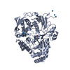
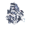
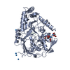
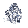
| Title | OLIGO-PEPTIDE BINDING PROTEIN (OPPA) COMPLEXED WITH KWK | ||||||
 Components Components |
| ||||||
 Keywords Keywords | COMPLEX (PEPTIDE TRANSPORT/PEPTIDE) / COMPLEX (PEPTIDE TRANSPORT-PEPTIDE) / PEPTIDE TRANSPORT / COMPLEX (PEPTIDE TRANSPORT-PEPTIDE) complex | ||||||
| Function / homology |  Function and homology information Function and homology informationpeptide transport / peptide transmembrane transporter activity / ATP-binding cassette (ABC) transporter complex / protein transport / outer membrane-bounded periplasmic space Similarity search - Function | ||||||
| Biological species |  Salmonella typhimurium (bacteria) Salmonella typhimurium (bacteria) | ||||||
| Method |  X-RAY DIFFRACTION / SYNCHROTRON / Resolution: 1.3 Å X-RAY DIFFRACTION / SYNCHROTRON / Resolution: 1.3 Å | ||||||
 Authors Authors | Tame, J. / Wilkinson, A.J. | ||||||
 Citation Citation | Journal: Nat.Struct.Biol. / Year: 1996 Title: The role of water in sequence-independent ligand binding by an oligopeptide transporter protein. Authors: Tame, J.R. / Sleigh, S.H. / Wilkinson, A.J. / Ladbury, J.E. #1: Journal: Structure / Year: 1995Title: The Crystal Structures of the Oligopeptide-Binding Protein Oppa Complexed with Tripeptide and Tetrapeptide Ligands Authors: Tame, J.R. / Dodson, E.J. / Murshudov, G. / Higgins, C.F. / Wilkinson, A.J. #2: Journal: Acta Crystallogr.,Sect.D / Year: 1995Title: Structure Determination of Oppa at 2.3 Angstroms Resolution Using Multiple Wavelength Anomalous Methods Authors: Glover, I.D. / Denny, R. / Nguti, N.D. / Mcsweeney, S. / Thompson, A. / Dodson, E. / Wilkinson, A.J. / Tame, J.R.H. #3: Journal: Science / Year: 1994Title: The Structural Basis of Sequence-Independent Peptide Binding by Oppa Protein Authors: Tame, J.R. / Murshudov, G.N. / Dodson, E.J. / Neil, T.K. / Dodson, G.G. / Higgins, C.F. / Wilkinson, A.J. | ||||||
| History |
|
- Structure visualization
| Structure viewer | Molecule: MolmilJmol/JSmol |
|---|
- Downloads & links
-Download
| PDBx/mmCIF format | 1jev.cif.gz | 128.1 KB | Display | PDBx/mmCIF format |
|---|---|---|---|---|
| PDB format | pdb1jev.ent.gz | 97.6 KB | Display | PDB format |
| PDBx/mmJSON format | 1jev.json.gz | Tree view | PDBx/mmJSON format | |
| Others |  Other downloads Other downloads |
-Validation report
| Arichive directory | https://data.pdbj.org/pub/pdb/validation_reports/je/1jevftp://data.pdbj.org/pub/pdb/validation_reports/je/1jev | HTTPS FTP |
|---|
-Related structure data

-Links
 PDBj
PDBj
- Assembly
| Deposited unit | 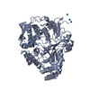
| ||||||||
|---|---|---|---|---|---|---|---|---|---|
| 1 |
| ||||||||
| Unit cell |
|
-Components
| #1: Protein | Mass: 58878.984 Da / Num. of mol.: 1 / Source method: isolated from a natural source / Source: (natural) Salmonella typhimurium (bacteria) / References: UniProt: P06202 | ||||
|---|---|---|---|---|---|
| #2: Protein/peptide | Mass: 462.585 Da / Num. of mol.: 1 Source method: isolated from a genetically manipulated source | ||||
| #3: Chemical | ChemComp-IUM /   Mass: 270.028 Da / Num. of mol.: 8 / Source method: obtained synthetically / Formula: O2U Mass: 270.028 Da / Num. of mol.: 8 / Source method: obtained synthetically / Formula: O2U#4: Water | ChemComp-HOH / |  Mass: 18.015 Da / Num. of mol.: 521 / Source method: isolated from a natural source / Formula: H2O Mass: 18.015 Da / Num. of mol.: 521 / Source method: isolated from a natural source / Formula: H2OHas protein modification | Y | |
-Experimental details
-Experiment
| Experiment | Method: X-RAY DIFFRACTION / Number of used crystals: 1 |
|---|
- Sample preparation
| Crystal | Density Matthews: 2.45 Å3/Da / Density % sol: 45 % / Description: FLASH COOLED TO 120K | ||||||||||||||||||||||||
|---|---|---|---|---|---|---|---|---|---|---|---|---|---|---|---|---|---|---|---|---|---|---|---|---|---|
| Crystal grow | pH: 5.5 / Details: CO-CRYSTALLIZED WITH URANIUM ACETATE, PH 5.5 | ||||||||||||||||||||||||
| Crystal grow | *PLUS Method: unknown / Details: Tame, J.R., (1994) Science, 264, 1578. | ||||||||||||||||||||||||
| Components of the solutions | *PLUS
|
-Data collection
| Diffraction | Mean temperature: 120 K |
|---|---|
| Diffraction source | Source: SYNCHROTRON / Site: EMBL/DESY, HAMBURG  / Beamline: X31 / Wavelength: 0.93 / Beamline: X31 / Wavelength: 0.93 |
| Detector | Type: MARRESEARCH / Detector: IMAGE PLATE |
| Radiation | Monochromatic (M) / Laue (L): M / Scattering type: x-ray |
| Radiation wavelength | Wavelength: 0.93 Å / Relative weight: 1 |
| Reflection | Num. obs: 143421 / % possible obs: 98.7 % / Observed criterion σ(I): 0 / Redundancy: 3.3 % / Biso Wilson estimate: 11.8 Å2 / Rmerge(I) obs: 0.035 |
| Reflection | *PLUS Highest resolution: 1.3 Å / Lowest resolution: 10 Å |
| Reflection shell | *PLUS % possible obs: 97.1 % |
- Processing
| Software |
| ||||||||||||||||||||||||||||||||||||||||||||||||||||||||||||||||||||||||||||||||||||
|---|---|---|---|---|---|---|---|---|---|---|---|---|---|---|---|---|---|---|---|---|---|---|---|---|---|---|---|---|---|---|---|---|---|---|---|---|---|---|---|---|---|---|---|---|---|---|---|---|---|---|---|---|---|---|---|---|---|---|---|---|---|---|---|---|---|---|---|---|---|---|---|---|---|---|---|---|---|---|---|---|---|---|---|---|---|
| Refinement | Resolution: 1.3→10 Å / σ(F): 0
| ||||||||||||||||||||||||||||||||||||||||||||||||||||||||||||||||||||||||||||||||||||
| Displacement parameters | Biso mean: 14.8 Å2 | ||||||||||||||||||||||||||||||||||||||||||||||||||||||||||||||||||||||||||||||||||||
| Refinement step | Cycle: LAST / Resolution: 1.3→10 Å
| ||||||||||||||||||||||||||||||||||||||||||||||||||||||||||||||||||||||||||||||||||||
| Refine LS restraints |
| ||||||||||||||||||||||||||||||||||||||||||||||||||||||||||||||||||||||||||||||||||||
| Software | *PLUS Name: PROLSQ / Classification: refinement | ||||||||||||||||||||||||||||||||||||||||||||||||||||||||||||||||||||||||||||||||||||
| Refinement | *PLUS Rfactor obs: 0.203 | ||||||||||||||||||||||||||||||||||||||||||||||||||||||||||||||||||||||||||||||||||||
| Solvent computation | *PLUS | ||||||||||||||||||||||||||||||||||||||||||||||||||||||||||||||||||||||||||||||||||||
| Displacement parameters | *PLUS | ||||||||||||||||||||||||||||||||||||||||||||||||||||||||||||||||||||||||||||||||||||
| LS refinement shell | *PLUS Rfactor Rfree: 0.28 / Rfactor obs: 0.29 |
