Protocol: SINGLE WAVELENGTH / Monochromatic (M) / Laue (L): M / Scattering type: x-ray
Radiation wavelength
Wavelength: 0.972 Å / Relative weight: 1
Reflection
Resolution: 2→50 Å / Num. obs: 345441 / % possible obs: 98.9 % / Observed criterion σ(I): -3 / Biso Wilson estimate: 32.543 Å2 / Rmerge(I) obs: 0.079 / Net I/σ(I): 12.41
Reflection shell
Resolution (Å)
Rmerge(I) obs
Mean I/σ(I) obs
Num. measured obs
Num. unique obs
Diffraction-ID
% possible all
2-2.12
0.554
2.26
183377
53085
1
94.2
2.12-2.26
0.354
3.69
195296
52829
1
99.7
2.26-2.44
0.259
5.07
187005
49270
1
99.9
2.44-2.68
0.173
7.43
174099
45338
1
99.9
2.68-2.99
0.106
11.59
158919
41163
1
99.9
2.99-3.45
0.061
19.22
139758
36189
1
99.9
3.45-4.22
0.039
29.61
117861
30701
1
99.9
4.22-5.95
0.031
35.27
91622
23801
1
99.9
-
Processing
Software
Name
Version
Classification
NB
XSCALE
datascaling
REFMAC
refinement
PDB_EXTRACT
3.1
dataextraction
Refinement
Method to determine structure: MOLECULAR REPLACEMENT / Resolution: 2→48.37 Å / Cor.coef. Fo:Fc: 0.922 / Cor.coef. Fo:Fc free: 0.897 / WRfactor Rfree: 0.2303 / WRfactor Rwork: 0.199 / Occupancy max: 1 / Occupancy min: 0.21 / FOM work R set: 0.8101 / SU B: 10.022 / SU ML: 0.15 / SU R Cruickshank DPI: 0.1979 / SU Rfree: 0.1719 / Cross valid method: THROUGHOUT / σ(F): 0 / ESU R Free: 0.179 / Stereochemistry target values: MAXIMUM LIKELIHOOD Details: HYDROGENS HAVE BEEN USED IF PRESENT IN THE INPUT U VALUES: WITH TLS ADDED
Rfactor
Num. reflection
% reflection
Selection details
Rfree
0.2666
17271
5 %
RANDOM
Rwork
0.2287
-
-
-
obs
0.2306
345407
98.87 %
-
Solvent computation
Ion probe radii: 0.8 Å / Shrinkage radii: 0.8 Å / VDW probe radii: 1.2 Å / Solvent model: MASK
In the structure databanks used in Yorodumi, some data are registered as the other names, "COVID-19 virus" and "2019-nCoV". Here are the details of the virus and the list of structure data.
Jan 31, 2019. EMDB accession codes are about to change! (news from PDBe EMDB page)
EMDB accession codes are about to change! (news from PDBe EMDB page)
The allocation of 4 digits for EMDB accession codes will soon come to an end. Whilst these codes will remain in use, new EMDB accession codes will include an additional digit and will expand incrementally as the available range of codes is exhausted. The current 4-digit format prefixed with “EMD-” (i.e. EMD-XXXX) will advance to a 5-digit format (i.e. EMD-XXXXX), and so on. It is currently estimated that the 4-digit codes will be depleted around Spring 2019, at which point the 5-digit format will come into force.
The EM Navigator/Yorodumi systems omit the EMD- prefix.
Related info.:Q: What is EMD? / ID/Accession-code notation in Yorodumi/EM Navigator
Yorodumi is a browser for structure data from EMDB, PDB, SASBDB, etc.
This page is also the successor to EM Navigator detail page, and also detail information page/front-end page for Omokage search.
The word "yorodu" (or yorozu) is an old Japanese word meaning "ten thousand". "mi" (miru) is to see.
Related info.:EMDB / PDB / SASBDB / Comparison of 3 databanks / Yorodumi Search / Aug 31, 2016. New EM Navigator & Yorodumi / Yorodumi Papers / Jmol/JSmol / Function and homology information / Changes in new EM Navigator and Yorodumi
 Movie
Movie Controller

 Yorodumi
 Open data
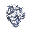
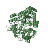
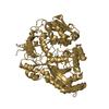
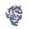
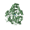
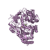
 Basic information
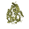
 Components
 Keywords
 Function and homology information
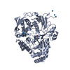
 X-RAY DIFFRACTION /
  Authors
 Citation
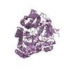
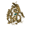
 Structure visualization
 Downloads & links
 Other downloads

 PDBj
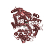
PDBj Assembly
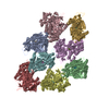


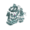
 Mass: 18.015 Da / Num. of mol.: 2408 / Source method: isolated from a natural source / Formula: H2O
 Sample preparation
 / Beamline: BW7A / Wavelength: 0.972 Å
 Processing