 Movie
Movie Controller

[English] 日本語
 Yorodumi
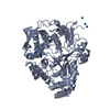
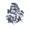
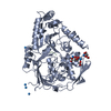
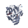
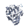
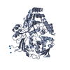
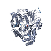
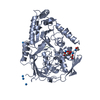
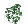
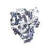
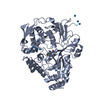
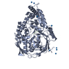
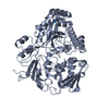
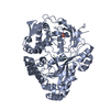
- PDB-1olc: OLIGO-PEPTIDE BINDING PROTEIN (OPPA) COMPLEXED WITH LYS-LYS-LYS-ALA -
+ Open data

- Basic information
| Entry | Database: PDB / ID: 1olc | ||||||
|---|---|---|---|---|---|---|---|
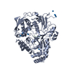
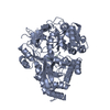
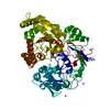
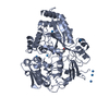
| Title | OLIGO-PEPTIDE BINDING PROTEIN (OPPA) COMPLEXED WITH LYS-LYS-LYS-ALA | ||||||
 Components Components |
| ||||||
 Keywords Keywords | COMPLEX (BINDING PROTEIN/PEPTIDE) / PERIPLASMIC / COMPLEX (BINDING PROTEIN-PEPTIDE) COMPLEX | ||||||
| Function / homology |  Function and homology information Function and homology informationpeptide transport / peptide transmembrane transporter activity / ATP-binding cassette (ABC) transporter complex / protein transport / outer membrane-bounded periplasmic space Similarity search - Function | ||||||
| Biological species |  Salmonella typhimurium (bacteria) Salmonella typhimurium (bacteria) | ||||||
| Method |  X-RAY DIFFRACTION / Resolution: 2.1 Å X-RAY DIFFRACTION / Resolution: 2.1 Å | ||||||
 Authors Authors | Tame, J. / Wilkinson, A.J. | ||||||
 Citation Citation | Journal: Structure / Year: 1995 Title: The crystal structures of the oligopeptide-binding protein OppA complexed with tripeptide and tetrapeptide ligands. Authors: Tame, J.R. / Dodson, E.J. / Murshudov, G. / Higgins, C.F. / Wilkinson, A.J. #1: Journal: Acta Crystallogr.,Sect.D / Year: 1995Title: Structure Determination of Oppa at 2.3 Angstroms Resolution Using Multiple Wavelength Anomalous Methods Authors: Glover, I.D. / Denny, R. / Nguti, N.D. / Mcsweeney, S. / Thompson, A. / Dodson, E. / Wilkinson, A.J. / Tame, J.R.H. #2: Journal: Science / Year: 1994Title: The Structural Basis of Sequence-Independent Peptide Binding by Oppa Protein Authors: Tame, J.R.H. / Murshudov, G. / Neil, T. / Dodson, E. / Dodson, G.G. / Higgins, C.F. / Wilkinson, A.J. | ||||||
| History |
| ||||||
| Remark 700 | SHEET THE HELIX AND SHEET RECORDS PRESENTED HERE ARE SIMPLIFIED TO HELP NAVIGATE AROUND THE PROTEIN ...SHEET THE HELIX AND SHEET RECORDS PRESENTED HERE ARE SIMPLIFIED TO HELP NAVIGATE AROUND THE PROTEIN AND TO AVOID OBSCURING TOPOLOGICAL RELATIONSHIPS WITH RELATED PROTEINS. IT IS NOT INTENDED TO REPLACE THE LIST WHICH THE PDB HAS GENERATED USING DSSP WHICH APPEAR ON ACTUAL HELIX AND SHEET RECORDS FURTHER DOWN IN THE ENTRY. BECAUSE OF LINE LENGTH LIMITATIONS THE FORMAT OF THE SHEET INFORMATION PRESENTED IN THIS REMARK HAS BEEN MODIFIED. HELIX 1 1 VAL A 34 LEU A 43 1 HELIX 2 2 HIS A 91 ALA A 101 1 HELIX 3 3 TYR A 112 GLY A 116 1 HELIX 4 4 ILE A 121 ALA A 126 1 HELIX 5 5 LYS A 169 PHE A 175 1 HELIX 6 6 GLU A 229 SER A 238 1 HELIX 7 7 ILE A 250 GLU A 259 1 HELIX 8 8 VAL A 287 ALA A 296 1 HELIX 9 9 ARG A 299 LYS A 305 1 HELIX 10 10 GLN A 337 ALA A 351 1 HELIX 11 11 ASP A 369 LEU A 386 1 HELIX 12 12 TRP A 397 GLN A 406 1 HELIX 13 13 THR A 424 LEU A 427 1 HELIX 14 14 PRO A 444 LYS A 455 1 HELIX 15 15 ASP A 459 ASP A 476 1 SH 1 A 7 VAL A 264 PRO A 268 0 SH 2 A 7 VAL A 486 LEU A 490 -1 N ARG A 489 O ARG A 265 SH 3 A 7 ASP A 242 TYR A 245 -1 N THR A 244 O LEU A 490 SH 4 A 7 THR A 14 ASN A 18 1 N ASN A 18 O MET A 243 SH 5 A 7 GLN A 220 LEU A 224 1 N GLN A 220 O LEU A 15 SH 6 A 7 ARG A 201 ARG A 206 -1 N LEU A 204 O VAL A 221 SH 7 A 7 TYR A 191 VAL A 197 -1 N VAL A 197 O ARG A 201 SH 1 B 4 ALA A 61 LYS A 67 0 SH 2 B 4 VAL A 71 LEU A 76 -1 N HIS A 75 O GLU A 62 SH 3 B 4 THR A 143 THR A 147 -1 N VAL A 146 O TRP A 72 SH 4 B 4 VAL A 136 ASP A 140 -1 N ASP A 140 O THR A 143 SH 1 C 5 ASN A 389 GLN A 395 0 SH 2 C 5 THR A 360 ASN A 366 1 O TYR A 365 N GLN A 395 SH 3 C 5 VAL A 411 CYS A 417 1 O VAL A 411 N LEU A 364 SH 4 C 5 CYS A 271 ILE A 277 -1 N GLU A 276 O ALA A 412 SH 5 C 5 ILE A 479 TYR A 484 -1 N TYR A 483 O TYR A 273 |
- Structure visualization
| Structure viewer | Molecule: MolmilJmol/JSmol |
|---|
- Downloads & links
-Download
| PDBx/mmCIF format | 1olc.cif.gz | 125 KB | Display | PDBx/mmCIF format |
|---|---|---|---|---|
| PDB format | pdb1olc.ent.gz | 95.7 KB | Display | PDB format |
| PDBx/mmJSON format | 1olc.json.gz | Tree view | PDBx/mmJSON format | |
| Others |  Other downloads Other downloads |
-Validation report
| Arichive directory | https://data.pdbj.org/pub/pdb/validation_reports/ol/1olcftp://data.pdbj.org/pub/pdb/validation_reports/ol/1olc | HTTPS FTP |
|---|
-Related structure data


-Links
 PDBj
PDBj
- Assembly
| Deposited unit | 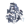
| ||||||||
|---|---|---|---|---|---|---|---|---|---|
| 1 |
| ||||||||
| Unit cell |
| ||||||||
| Atom site foot note | 1: CIS PROLINE - PRO A 283 |
-Components
| #1: Protein | Mass: 58878.984 Da / Num. of mol.: 1 / Source method: isolated from a natural source / Source: (natural) Salmonella typhimurium (bacteria) / References: UniProt: P06202 | ||||
|---|---|---|---|---|---|
| #2: Protein/peptide | Mass: 476.632 Da / Num. of mol.: 1 Source method: isolated from a genetically manipulated source Details: CO-CRYSTALLIZED WITH URANIUM ACETATE | ||||
| #3: Chemical | ChemComp-IUM /   Mass: 270.028 Da / Num. of mol.: 8 / Source method: obtained synthetically / Formula: O2U Mass: 270.028 Da / Num. of mol.: 8 / Source method: obtained synthetically / Formula: O2U#4: Water | ChemComp-HOH / |  Mass: 18.015 Da / Num. of mol.: 337 / Source method: isolated from a natural source / Formula: H2O Mass: 18.015 Da / Num. of mol.: 337 / Source method: isolated from a natural source / Formula: H2OHas protein modification | Y | |
-Experimental details
-Experiment
| Experiment | Method: X-RAY DIFFRACTION / Number of used crystals: 1 |
|---|
- Sample preparation
| Crystal | Density Matthews: 2.56 Å3/Da / Density % sol: 52.01 % | ||||||||||||||||||||
|---|---|---|---|---|---|---|---|---|---|---|---|---|---|---|---|---|---|---|---|---|---|
| Crystal grow | pH: 5.5 / Details: pH 5.5 | ||||||||||||||||||||
| Crystal grow | *PLUS Method: unknown | ||||||||||||||||||||
| Components of the solutions | *PLUS
|
-Data collection
| Diffraction | Mean temperature: 291 K |
|---|---|
| Diffraction source | Wavelength: 1.5418 Å |
| Detector | Type: RIGAKU RAXIS IIC / Detector: IMAGE PLATE |
| Radiation | Monochromatic (M) / Laue (L): M / Scattering type: x-ray |
| Radiation wavelength | Wavelength: 1.5418 Å / Relative weight: 1 |
| Reflection | Resolution: 2→20 Å / Num. obs: 41810 / % possible obs: 99.7 % / Observed criterion σ(I): 0 / Redundancy: 4.7 % / Rmerge(I) obs: 0.063 |
| Reflection | *PLUS Rmerge(I) obs: 0.063 |
| Reflection shell | *PLUS % possible obs: 100 % / Rmerge(I) obs: 0.176 / Mean I/σ(I) obs: 3.8 |
- Processing
| Software |
| ||||||||||||||||||||||||||||||||||||||||||||||||||||||||||||||||||||||||||||||||||||
|---|---|---|---|---|---|---|---|---|---|---|---|---|---|---|---|---|---|---|---|---|---|---|---|---|---|---|---|---|---|---|---|---|---|---|---|---|---|---|---|---|---|---|---|---|---|---|---|---|---|---|---|---|---|---|---|---|---|---|---|---|---|---|---|---|---|---|---|---|---|---|---|---|---|---|---|---|---|---|---|---|---|---|---|---|---|
| Refinement | Resolution: 2.1→10 Å / σ(F): 0
| ||||||||||||||||||||||||||||||||||||||||||||||||||||||||||||||||||||||||||||||||||||
| Displacement parameters | Biso mean: 20.3 Å2 | ||||||||||||||||||||||||||||||||||||||||||||||||||||||||||||||||||||||||||||||||||||
| Refinement step | Cycle: LAST / Resolution: 2.1→10 Å
| ||||||||||||||||||||||||||||||||||||||||||||||||||||||||||||||||||||||||||||||||||||
| Refine LS restraints |
|
