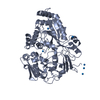
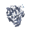
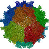
PDB-9rdu F001 手法 : X-RAY DIFFRACTION / 解像度 : 1.42 Å
PDB-9rdv F005 手法 : X-RAY DIFFRACTION / 解像度 : 1.32 Å
PDB-9rdw F009 手法 : X-RAY DIFFRACTION / 解像度 : 1.44 Å
PDB-9rdx F012 手法 : X-RAY DIFFRACTION / 解像度 : 1.5 Å
PDB-9rdy F024 手法 : X-RAY DIFFRACTION / 解像度 : 1.5 Å
PDB-9rdz F030 手法 : X-RAY DIFFRACTION / 解像度 : 1.64 Å
PDB-9re0 F032 手法 : X-RAY DIFFRACTION / 解像度 : 1.58 Å
PDB-9re1 F055 手法 : X-RAY DIFFRACTION / 解像度 : 1.41 Å
PDB-9re2 F058 手法 : X-RAY DIFFRACTION / 解像度 : 1.68 Å
PDB-9re3 F070 手法 : X-RAY DIFFRACTION / 解像度 : 1.57 Å
PDB-9re4 F073 手法 : X-RAY DIFFRACTION / 解像度 : 1.47 Å
PDB-9re5 F074 手法 : X-RAY DIFFRACTION / 解像度 : 1.7 Å
PDB-9re6 F102 手法 : X-RAY DIFFRACTION / 解像度 : 1.45 Å
PDB-9re7 F134 手法 : X-RAY DIFFRACTION / 解像度 : 1.46 Å
PDB-9re8 F138 手法 : X-RAY DIFFRACTION / 解像度 : 1.5 Å
PDB-9re9 F145 手法 : X-RAY DIFFRACTION / 解像度 : 1.41 Å
PDB-9rea F168 手法 : X-RAY DIFFRACTION / 解像度 : 1.52 Å
PDB-9reb F184 手法 : X-RAY DIFFRACTION / 解像度 : 1.39 Å
PDB-9rec F186 手法 : X-RAY DIFFRACTION / 解像度 : 1.2 Å
PDB-9red F188 手法 : X-RAY DIFFRACTION / 解像度 : 1.31 Å
PDB-9ree F189 手法 : X-RAY DIFFRACTION / 解像度 : 1.34 Å
PDB-9ref F203 手法 : X-RAY DIFFRACTION / 解像度 : 1.42 Å
PDB-9reg F225 手法 : X-RAY DIFFRACTION / 解像度 : 1.35 Å
PDB-9reh F236 手法 : X-RAY DIFFRACTION / 解像度 : 1.33 Å
PDB-9rei F248 手法 : X-RAY DIFFRACTION / 解像度 : 1.42 Å
PDB-9rej F264 手法 : X-RAY DIFFRACTION / 解像度 : 1.4 Å
PDB-9rek F274 手法 : X-RAY DIFFRACTION / 解像度 : 1.38 Å
PDB-9rel F283 手法 : X-RAY DIFFRACTION / 解像度 : 1.42 Å
PDB-9rem F294 手法 : X-RAY DIFFRACTION / 解像度 : 1.43 Å
PDB-9ren F296 手法 : X-RAY DIFFRACTION / 解像度 : 1.38 Å
PDB-9reo F299 手法 : X-RAY DIFFRACTION / 解像度 : 1.45 Å
PDB-9rep F304 手法 : X-RAY DIFFRACTION / 解像度 : 1.44 Å
PDB-9req F310 手法 : X-RAY DIFFRACTION / 解像度 : 1.5 Å
PDB-9rer F312 手法 : X-RAY DIFFRACTION / 解像度 : 1.45 Å
PDB-9res F313 手法 : X-RAY DIFFRACTION / 解像度 : 1.5 Å
PDB-9ret F322 手法 : X-RAY DIFFRACTION / 解像度 : 1.75 Å
 ムービー
ムービー コントローラー
 構造ビューア
 万見文献について


 著者
著者 リンク
リンク


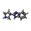


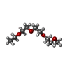


 キーワード
キーワード
 cricetulus griseus (モンゴルキヌゲネズミ)