 ムービー
ムービー コントローラー
 構造ビューア
 EMN検索について


-検索条件
-検索結果
検索 (著者・登録者: lill & p)の結果全44件を表示しています
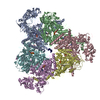
EMDB-17731: 
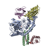
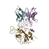
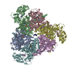
Structure of the human mitochondrial iron-sulfur cluster biosynthesis complex during persulfide transfer (consensus map)
EMDB-17732:
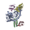
Structure of the human mitochondrial iron-sulfur cluster biosynthesis complex during persulfide transfer (persulfide on ISCU2)
EMDB-17733:
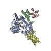
Structure of the human mitochondrial iron-sulfur cluster biosynthesis complex during persulfide transfer (persulfide on NFS1 and ISCU2)
EMDB-17734:
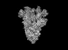
Structure of the human mitochondrial iron-sulfur cluster biosynthesis complex during persulfide transfer (without frataxin)
PDB-8pk8:
Structure of the human mitochondrial iron-sulfur cluster biosynthesis complex during persulfide transfer (persulfide on ISCU2)
PDB-8pk9:
Structure of the human mitochondrial iron-sulfur cluster biosynthesis complex during persulfide transfer (persulfide on NFS1 and ISCU2)
PDB-8pka:
Structure of the human mitochondrial iron-sulfur cluster biosynthesis complex during persulfide transfer (without frataxin)
EMDB-28198:
Cryo-EM map of SARS-CoV-2 Omicron BA.2 spike in complex with LLNL-199

EMDB-28199:
Cryo-EM map of SARS-CoV-2 Omicron BA.2 spike in complex with 2130-1-0114-112
PDB-8ekd:
Cryo-EM map of SARS-CoV-2 Omicron BA.2 spike in complex with 2130-1-0114-112
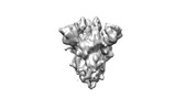
EMDB-16372:
Structure of the peroxisomal Pex1/Pex6 ATPase complex bound to a substrate in single seam state
EMDB-16373:
Structure of the peroxisomal Pex1/Pex6 ATPase complex bound to a substrate in twin seam state
PDB-8c0v:
Structure of the peroxisomal Pex1/Pex6 ATPase complex bound to a substrate in single seam state
PDB-8c0w:
Structure of the peroxisomal Pex1/Pex6 ATPase complex bound to a substrate in twin seam state

EMDB-15646:
Wild type hexamer oxalyl-CoA synthetase (OCS)

PDB-8atd:
Wild type hexamer oxalyl-CoA synthetase (OCS)

EMDB-29052:
SARS-CoV-2 Spike Hexapro - C68.59 Fab (Class 3 - disordered)
EMDB-29053:
SARS-CoV-2 Spike Hexapro - C59.68 Fab (Class 1 - No Fab bound)

EMDB-29054:
SARS-CoV-2 Spike Hexapro - C68.59 Fab (Class 2 - Fab bound)

EMDB-26593:
CryoEM structure of full-length dimeric ClbP

PDB-7ul6:
CryoEM structure of full-length dimeric ClbP

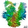
EMDB-33152:
Structural basis for Gemin5 decamer-mediated mRNA binding

EMDB-33187:
Structure of Gemin5 C-terminal region (protomer)

PDB-7xdt:
Structural basis for Gemin5 decamer-mediated mRNA binding

PDB-7xgr:
Structure of Gemin5 C-terminal region (protomer)

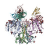
EMDB-23462:
Structure of CD4 mimetic BNM-III-170 in complex with BG505 SOSIP.664 HIV-1 Env trimer and 17b Fab
EMDB-23465:
Structure of CD4 mimetic M48U1 in complex with BG505 SOSIP.664 HIV-1 Env trimer and 17b Fab
PDB-7lo6:
Structure of CD4 mimetic BNM-III-170 in complex with BG505 SOSIP.664 HIV-1 Env trimer and 17b Fab

PDB-7lok:
Structure of CD4 mimetic M48U1 in complex with BG505 SOSIP.664 HIV-1 Env trimer and 17b Fab
EMDB-12047:
CryoEM Structure of the yeast peroxisomal membrane Pex14p/Pex17p complex
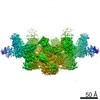
EMDB-21226:
Negative stain reconstruction of the yeast exocyst octameric complex.
PDB-6vkl:
Negative stain reconstruction of the yeast exocyst octameric complex.

EMDB-21132:
Cryo-EM structure of PCAT1 bound to its CtA peptide substrate

PDB-6v9z:
Cryo-EM structure of PCAT1 bound to its CtA peptide substrate
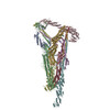
EMDB-0649:
Electron cryo-microscopy of the eukaryotic translation initiation factor 2B bound to translation initiation factor 2 from Homo sapiens
EMDB-0651:
Electron cryo-microscopy of the eukaryotic translation initiation factor 2B bound to eukaryotic translation initiation factor 2 from Homo sapiens
EMDB-0664:
Electron cryo-microscopy of the eukaryotic translation initiation factor 2B bound to eukaryotic translation initiation factor 2 from Homo sapiens
PDB-6o81:
Electron cryo-microscopy of the eukaryotic translation initiation factor 2B bound to translation initiation factor 2 from Homo sapiens

PDB-6o85:
Electron cryo-microscopy of the eukaryotic translation initiation factor 2B bound to eukaryotic translation initiation factor 2 from Homo sapiens

PDB-6o9z:
Electron cryo-microscopy of the eukaryotic translation initiation factor 2B bound to eukaryotic translation initiation factor 2 from Homo sapiens

EMDB-0466:
HIV-1 Env State 2A
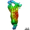
EMDB-3809:
The Cryo-Electron Microscopy Structure of the Type 1 Chaperone-Usher Pilus Rod

PDB-5oh0:
The Cryo-Electron Microscopy Structure of the Type 1 Chaperone-Usher Pilus Rod
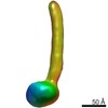
EMDB-3391:
Negative-stain electron microscopy structure of human cytomegalovirus gHgLgO trimer
 wwPDBはEMDBデータモデルのバージョン3へ移行します
