[English] 日本語
 Yorodumi
- EMDB-3391: Negative-stain electron microscopy structure of human cytomegalov... -
+ Open data
- Basic information
| Entry | Database: EMDB / ID: EMD-3391 | |||||||||
|---|---|---|---|---|---|---|---|---|---|---|
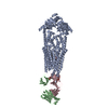
| Title | Negative-stain electron microscopy structure of human cytomegalovirus gHgLgO trimer | |||||||||
 Map data Map data | The correct handedness for this complex was unknown at the time of deposition | |||||||||
 Sample Sample |
| |||||||||
 Keywords Keywords | gHgLgO / human cytomegalovirus | |||||||||
| Function / homology |  Function and homology information Function and homology informationviral process / membrane => GO:0016020 / host cell endosome / host cell Golgi apparatus / entry receptor-mediated virion attachment to host cell / fusion of virus membrane with host plasma membrane / viral envelope / symbiont entry into host cell / host cell plasma membrane / virion membrane / membrane Similarity search - Function | |||||||||
| Biological species |   Human herpesvirus 5 Human herpesvirus 5 | |||||||||
| Method | single particle reconstruction / negative staining / Resolution: 19.3 Å | |||||||||
 Authors Authors | Kabanova A / Marcandalli J / Zhou T / Bianchi S / Baxa U / Tsybovsky Y / Lilleri D / Silacci-Fregni C / Foglierini M / Fernandez-Rodriguez BM ...Kabanova A / Marcandalli J / Zhou T / Bianchi S / Baxa U / Tsybovsky Y / Lilleri D / Silacci-Fregni C / Foglierini M / Fernandez-Rodriguez BM / Druz A / Zhang B / Geiger R / Pagani M / Sallusto F / Kwong PD / Corti D / Lanzavecchia A / Perez L | |||||||||
 Citation Citation |  Journal: Nat Microbiol / Year: 2016 Journal: Nat Microbiol / Year: 2016Title: Platelet-derived growth factor-α receptor is the cellular receptor for human cytomegalovirus gHgLgO trimer. Authors: Anna Kabanova / Jessica Marcandalli / Tongqing Zhou / Siro Bianchi / Ulrich Baxa / Yaroslav Tsybovsky / Daniele Lilleri / Chiara Silacci-Fregni / Mathilde Foglierini / Blanca Maria Fernandez- ...Authors: Anna Kabanova / Jessica Marcandalli / Tongqing Zhou / Siro Bianchi / Ulrich Baxa / Yaroslav Tsybovsky / Daniele Lilleri / Chiara Silacci-Fregni / Mathilde Foglierini / Blanca Maria Fernandez-Rodriguez / Aliaksandr Druz / Baoshan Zhang / Roger Geiger / Massimiliano Pagani / Federica Sallusto / Peter D Kwong / Davide Corti / Antonio Lanzavecchia / Laurent Perez /    Abstract: Human cytomegalovirus encodes at least 25 membrane glycoproteins that are found in the viral envelope(1). While gB represents the fusion protein, two glycoprotein complexes control the tropism of the ...Human cytomegalovirus encodes at least 25 membrane glycoproteins that are found in the viral envelope(1). While gB represents the fusion protein, two glycoprotein complexes control the tropism of the virus: the gHgLgO trimer is involved in the infection of fibroblasts, and the gHgLpUL128L pentamer is required for infection of endothelial, epithelial and myeloid cells(2-5). Two reports suggested that gB binds to ErbB1 and PDGFRα (refs 6,7); however, these results do not explain the tropism of the virus and were recently challenged(8,9). Here, we provide a 19 Å reconstruction for the gHgLgO trimer and show that it binds with high affinity through the gO subunit to PDGFRα, which is expressed on fibroblasts but not on epithelial cells. We also provide evidence that the trimer is essential for viral entry in both fibroblasts and epithelial cells. Furthermore, we identify the pentamer, which is essential for infection of epithelial cells, as a trigger for the ErbB pathway. These findings help explain the broad tropism of human cytomegalovirus and indicate that PDGFRα and the viral gO subunit could be targeted by novel anti-viral therapies. | |||||||||
| History |
|
- Structure visualization
| Movie |
 Movie viewer Movie viewer |
|---|---|
| Structure viewer | EM map:  SurfView SurfView Molmil Molmil Jmol/JSmol Jmol/JSmol |
| Supplemental images |
- Downloads & links
-EMDB archive
| Map data |  emd_3391.map.gz emd_3391.map.gz | 14.7 MB |  EMDB map data format EMDB map data format | |
|---|---|---|---|---|
| Header (meta data) |  emd-3391-v30.xml emd-3391-v30.xml emd-3391.xml emd-3391.xml | 13.6 KB 13.6 KB | Display Display |  EMDB header EMDB header |
| Images |  EMD-3391.tif EMD-3391.tif | 51.7 KB | ||
| Archive directory |  http://ftp.pdbj.org/pub/emdb/structures/EMD-3391 http://ftp.pdbj.org/pub/emdb/structures/EMD-3391 ftp://ftp.pdbj.org/pub/emdb/structures/EMD-3391 ftp://ftp.pdbj.org/pub/emdb/structures/EMD-3391 | HTTPS FTP |
-Validation report
| Summary document |  emd_3391_validation.pdf.gz emd_3391_validation.pdf.gz | 200.5 KB | Display |  EMDB validaton report EMDB validaton report |
|---|---|---|---|---|
| Full document |  emd_3391_full_validation.pdf.gz emd_3391_full_validation.pdf.gz | 199.6 KB | Display | |
| Data in XML |  emd_3391_validation.xml.gz emd_3391_validation.xml.gz | 5.7 KB | Display | |
| Arichive directory |  https://ftp.pdbj.org/pub/emdb/validation_reports/EMD-3391 https://ftp.pdbj.org/pub/emdb/validation_reports/EMD-3391 ftp://ftp.pdbj.org/pub/emdb/validation_reports/EMD-3391 ftp://ftp.pdbj.org/pub/emdb/validation_reports/EMD-3391 | HTTPS FTP |
-Related structure data
| Similar structure data |
|---|
- Links
Links
| EMDB pages |  EMDB (EBI/PDBe) / EMDB (EBI/PDBe) /  EMDataResource EMDataResource |
|---|
- Map
Map
| File |  Download / File: emd_3391.map.gz / Format: CCP4 / Size: 15.3 MB / Type: IMAGE STORED AS FLOATING POINT NUMBER (4 BYTES) Download / File: emd_3391.map.gz / Format: CCP4 / Size: 15.3 MB / Type: IMAGE STORED AS FLOATING POINT NUMBER (4 BYTES) | ||||||||||||||||||||||||||||||||||||||||||||||||||||||||||||
|---|---|---|---|---|---|---|---|---|---|---|---|---|---|---|---|---|---|---|---|---|---|---|---|---|---|---|---|---|---|---|---|---|---|---|---|---|---|---|---|---|---|---|---|---|---|---|---|---|---|---|---|---|---|---|---|---|---|---|---|---|---|
| Annotation | The correct handedness for this complex was unknown at the time of deposition | ||||||||||||||||||||||||||||||||||||||||||||||||||||||||||||
| Projections & slices | Image control
Images are generated by Spider. | ||||||||||||||||||||||||||||||||||||||||||||||||||||||||||||
| Voxel size | X=Y=Z: 2.22 Å | ||||||||||||||||||||||||||||||||||||||||||||||||||||||||||||
| Density |
| ||||||||||||||||||||||||||||||||||||||||||||||||||||||||||||
| Symmetry | Space group: 1 | ||||||||||||||||||||||||||||||||||||||||||||||||||||||||||||
| Details | EMDB XML:
CCP4 map header:
| ||||||||||||||||||||||||||||||||||||||||||||||||||||||||||||
-Supplemental data
- Sample components
-Entire : Human cytomegalovirus gHgLgO trimer
| Entire | Name: Human cytomegalovirus gHgLgO trimer |
|---|---|
| Components |
|
-Supramolecule #1000: Human cytomegalovirus gHgLgO trimer
| Supramolecule | Name: Human cytomegalovirus gHgLgO trimer / type: sample / ID: 1000 / Oligomeric state: Heterotrimer (one gH + one gL + one gO) / Number unique components: 3 |
|---|---|
| Molecular weight | Experimental: 390 KDa / Theoretical: 170 KDa / Method: Size exclusion chromatography |
-Macromolecule #1: Envelope glycoprotein H
| Macromolecule | Name: Envelope glycoprotein H / type: protein_or_peptide / ID: 1 / Name.synonym: gH, UL75 / Number of copies: 1 / Oligomeric state: Heterotrimeric with gL and gO / Recombinant expression: Yes |
|---|---|
| Source (natural) | Organism:   Human herpesvirus 5 / Strain: VR1814 / synonym: HCMV Human herpesvirus 5 / Strain: VR1814 / synonym: HCMV |
| Molecular weight | Theoretical: 85 KDa |
| Recombinant expression | Organism: Chinese Hamster Ovary cells / Recombinant cell: CHO K1SV / Recombinant plasmid: PEE.12.4 and PEE6.4 |
| Sequence | UniProtKB: Envelope glycoprotein H / GO: membrane, membrane => GO:0016020, viral envelope / InterPro: Herpesvirus glycoprotein H |
-Macromolecule #2: Envelope glycoprotein O
| Macromolecule | Name: Envelope glycoprotein O / type: protein_or_peptide / ID: 2 / Name.synonym: gO, UL74 / Number of copies: 1 / Oligomeric state: Heterotrimeric with gL and gH / Recombinant expression: Yes |
|---|---|
| Source (natural) | Organism:   Human herpesvirus 5 / Strain: VR1814 / synonym: HCMV Human herpesvirus 5 / Strain: VR1814 / synonym: HCMV |
| Molecular weight | Theoretical: 54 KDa |
| Recombinant expression | Organism: Chinese Hamster Ovary cells / Recombinant cell: CHO K1SV / Recombinant plasmid: PEE.12.4 and PEE6.4 |
| Sequence | UniProtKB: Envelope glycoprotein O / GO: viral envelope / InterPro: Herpesvirus UL74, glycoprotein |
-Macromolecule #3: Envelope glycoprotein L
| Macromolecule | Name: Envelope glycoprotein L / type: protein_or_peptide / ID: 3 / Name.synonym: gL, UL115 / Number of copies: 1 / Oligomeric state: Heterotrimeric with gH and gO / Recombinant expression: Yes |
|---|---|
| Source (natural) | Organism:   Human herpesvirus 5 / Strain: VR1814 / synonym: HCMV Human herpesvirus 5 / Strain: VR1814 / synonym: HCMV |
| Molecular weight | Theoretical: 30 KDa |
| Recombinant expression | Organism: Chinese Hamster Ovary cells / Recombinant cell: CHO K1SV / Recombinant plasmid: PEE.12.4 and PEE6.4 |
| Sequence | UniProtKB: Envelope glycoprotein L / GO: viral process, viral envelope / InterPro: Cytomegalovirus glycoprotein L |
-Experimental details
-Structure determination
| Method | negative staining |
|---|---|
 Processing Processing | single particle reconstruction |
| Aggregation state | particle |
- Sample preparation
| Concentration | 0.01 mg/mL |
|---|---|
| Buffer | pH: 7.5 / Details: 20 mM HEPES, 150 mM NaCl |
| Staining | Type: NEGATIVE Details: The complex was adsorbed to freshly glow-discharged carbon-coated grids for 15 s, washed with a buffer containing 20 mM HEPES, pH 7.5, and 150 mM NaCl, and stained in several drops of 0.7% uranyl formate |
| Grid | Details: 200 mesh copper grid with carbon support, glow discharged |
| Vitrification | Cryogen name: NONE / Instrument: OTHER |
- Electron microscopy
| Microscope | FEI TECNAI 20 |
|---|---|
| Alignment procedure | Legacy - Astigmatism: Objective lens astigmatism was corrected at 100,000 times magnification |
| Date | Jun 18, 2015 |
| Image recording | Category: CCD / Film or detector model: FEI EAGLE (2k x 2k) / Number real images: 304 |
| Electron beam | Acceleration voltage: 200 kV / Electron source: LAB6 |
| Electron optics | Illumination mode: FLOOD BEAM / Imaging mode: BRIGHT FIELD / Cs: 2.0 mm / Nominal defocus max: 0.9 µm / Nominal defocus min: 0.4 µm / Nominal magnification: 100000 |
| Sample stage | Specimen holder: Room temperature / Specimen holder model: SIDE ENTRY, EUCENTRIC |
- Image processing
| Details | The dataset was subjected to iterative stable alignment and clustering (ISAC) in SPARX. The ISAC classes were then used to generate an initial 3D map in EMAN2, which was used as the initial model for three-dimensional reconstruction and refinement using reference projections in SPIDER. |
|---|---|
| CTF correction | Details: Each defocus group |
| Final reconstruction | Applied symmetry - Point group: C1 (asymmetric) / Algorithm: OTHER / Resolution.type: BY AUTHOR / Resolution: 19.3 Å / Resolution method: OTHER / Software - Name: SPARX, EMAN2, SPIDER / Number images used: 10356 |
| Final two d classification | Number classes: 37 |
 Movie
Movie Controller


 UCSF Chimera


 Z (Sec.)
 Y (Row.)
 X (Col.)


