 Movie
Movie Controller

[English] 日本語
 Yorodumi
- PDB-5oh0: The Cryo-Electron Microscopy Structure of the Type 1 Chaperone-Us... -
+ Open data

- Basic information
| Entry | Database: PDB / ID: 5oh0 | ||||||
|---|---|---|---|---|---|---|---|
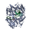
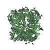
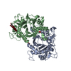
| Title | The Cryo-Electron Microscopy Structure of the Type 1 Chaperone-Usher Pilus Rod | ||||||
 Components Components | Type-1 fimbrial protein, A chain | ||||||
 Keywords Keywords | PROTEIN FIBRIL / bacterial pilus / chaperone-usher pilus | ||||||
| Function / homology |  Function and homology information Function and homology informationcell adhesion involved in single-species biofilm formation / pilus / cell adhesion / identical protein binding Similarity search - Function | ||||||
| Biological species |  | ||||||
| Method | ELECTRON MICROSCOPY / helical reconstruction / cryo EM / Resolution: 4.2 Å | ||||||
 Authors Authors | Hospenthal, M.K. / Costa, T.R.D. / Redzej, A. / Waksman, G. | ||||||
| Funding support |  United Kingdom, 1items United Kingdom, 1items
| ||||||
 Citation Citation | Journal: Structure / Year: 2017 Title: The Cryoelectron Microscopy Structure of the Type 1 Chaperone-Usher Pilus Rod. Authors: Manuela K Hospenthal / Dawid Zyla / Tiago R D Costa / Adam Redzej / Christoph Giese / James Lillington / Rudi Glockshuber / Gabriel Waksman /  Abstract: Adhesive chaperone-usher pili are long, supramolecular protein fibers displayed on the surface of many bacterial pathogens. The type 1 and P pili of uropathogenic Escherichia coli (UPEC) play ...Adhesive chaperone-usher pili are long, supramolecular protein fibers displayed on the surface of many bacterial pathogens. The type 1 and P pili of uropathogenic Escherichia coli (UPEC) play important roles during urinary tract colonization, mediating attachment to the bladder and kidney, respectively. The biomechanical properties of the helical pilus rods allow them to reversibly uncoil in response to flow-induced forces, allowing UPEC to retain a foothold in the unique and hostile environment of the urinary tract. Here we provide the 4.2-Å resolution cryo-EM structure of the type 1 pilus rod, which together with the previous P pilus rod structure rationalizes the remarkable "spring-like" properties of chaperone-usher pili. The cryo-EM structure of the type 1 pilus rod differs in its helical parameters from the structure determined previously by a hybrid approach. We provide evidence that these structural differences originate from different quaternary structures of pili assembled in vivo and in vitro. | ||||||
| History |
|
- Structure visualization
| Movie |
Movie viewer |
|---|---|
| Structure viewer | Molecule: MolmilJmol/JSmol |
- Downloads & links
-Download
| PDBx/mmCIF format | 5oh0.cif.gz | 149.3 KB | Display | PDBx/mmCIF format |
|---|---|---|---|---|
| PDB format | pdb5oh0.ent.gz | 119.7 KB | Display | PDB format |
| PDBx/mmJSON format | 5oh0.json.gz | Tree view | PDBx/mmJSON format | |
| Others |  Other downloads Other downloads |
-Validation report
| Arichive directory | https://data.pdbj.org/pub/pdb/validation_reports/oh/5oh0ftp://data.pdbj.org/pub/pdb/validation_reports/oh/5oh0 | HTTPS FTP |
|---|
-Related structure data
| Related structure data | 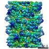 3809MC M: map data used to model this data C: citing same article ( |
|---|---|
| Similar structure data |


-Links
 PDBj
PDBj- Assembly
| Deposited unit | 
|
|---|---|
| 1 |
|
-Components
| #1: Protein | Mass: 15835.243 Da / Num. of mol.: 6 Source method: isolated from a genetically manipulated source Source: (gene. exp.) Has protein modification | Y | |
|---|
-Experimental details
-Experiment
| Experiment | Method: ELECTRON MICROSCOPY |
|---|---|
| EM experiment | Aggregation state: HELICAL ARRAY / 3D reconstruction method: helical reconstruction |
- Sample preparation
| Component | Name: Type 1 Chaperone-usher pilus / Type: COMPLEX Details: Superhelical assembly of the pilus rod subunit FimA Entity ID: all / Source: RECOMBINANT | ||||||||||||
|---|---|---|---|---|---|---|---|---|---|---|---|---|---|
| Molecular weight | Experimental value: NO | ||||||||||||
| Source (natural) | Organism: | ||||||||||||
| Source (recombinant) | Organism: | ||||||||||||
| Buffer solution | pH: 7.5 | ||||||||||||
| Buffer component |
| ||||||||||||
| Specimen | Embedding applied: NO / Shadowing applied: NO / Staining applied: NO / Vitrification applied: YES | ||||||||||||
| Specimen support | Grid type: Quantifoil 1.2/1.3 400 mesh grid | ||||||||||||
| Vitrification | Cryogen name: ETHANE / Humidity: 95 % |
- Electron microscopy imaging
| Experimental equipment |  Model: Tecnai Polara / Image courtesy: FEI Company |
|---|---|
| Microscopy | Model: FEI POLARA 300 |
| Electron gun | Electron source:  FIELD EMISSION GUN / Accelerating voltage: 300 kV / Illumination mode: FLOOD BEAM FIELD EMISSION GUN / Accelerating voltage: 300 kV / Illumination mode: FLOOD BEAM |
| Electron lens | Mode: BRIGHT FIELD |
| Image recording | Electron dose: 1.7 e/Å2 / Film or detector model: GATAN K2 SUMMIT (4k x 4k) |
- Processing
| Software | Name: PHENIX / Version: dev_2776: / Classification: refinement | |||||||||||||||||||||||||
|---|---|---|---|---|---|---|---|---|---|---|---|---|---|---|---|---|---|---|---|---|---|---|---|---|---|---|
| EM software |
| |||||||||||||||||||||||||
| CTF correction | Type: PHASE FLIPPING AND AMPLITUDE CORRECTION | |||||||||||||||||||||||||
| Helical symmerty | Angular rotation/subunit: 114.992 ° / Axial rise/subunit: 8.01188 Å / Axial symmetry: C1 | |||||||||||||||||||||||||
| Particle selection | Num. of particles selected: 115545 | |||||||||||||||||||||||||
| 3D reconstruction | Resolution: 4.2 Å / Resolution method: FSC 0.143 CUT-OFF / Num. of particles: 115510 / Symmetry type: HELICAL | |||||||||||||||||||||||||
| Atomic model building | 3D fitting-ID: 1 / Accession code: 2JTY / Initial refinement model-ID: 1 / Pdb chain-ID: A / PDB-ID: 2JTY / Source name: PDB / Type: experimental model
| |||||||||||||||||||||||||
| Refine LS restraints |
|

